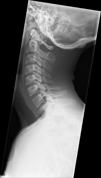
File:Normal trauma cervical spine (Radiopaedia 41017-43761 Lateral 1).png
Jump to navigation
Jump to search

Size of this preview: 341 × 599 pixels. Other resolutions: 136 × 240 pixels | 273 × 480 pixels | 437 × 768 pixels | 583 × 1,024 pixels | 1,456 × 2,556 pixels.
Original file (1,456 × 2,556 pixels, file size: 2.23 MB, MIME type: image/png)
Summary:
| Description |
|
| Date | Published: 18th Nov 2015 |
| Source | https://radiopaedia.org/cases/normal-trauma-cervical-spine |
| Author | Bruno Di Muzio |
| Permission (Permission-reusing-text) |
http://creativecommons.org/licenses/by-nc-sa/3.0/ |
Licensing:
Attribution-NonCommercial-ShareAlike 3.0 Unported (CC BY-NC-SA 3.0)
File history
Click on a date/time to view the file as it appeared at that time.
| Date/Time | Thumbnail | Dimensions | User | Comment | |
|---|---|---|---|---|---|
| current | 11:11, 28 August 2021 | | 1,456 × 2,556 (2.23 MB) | Fæ (talk | contribs) | Radiopaedia project rID:41017 (thread B) (batch #26705-1 A1) |
You cannot overwrite this file.
File usage
There are no pages that use this file.
.png&oldid=1170876)