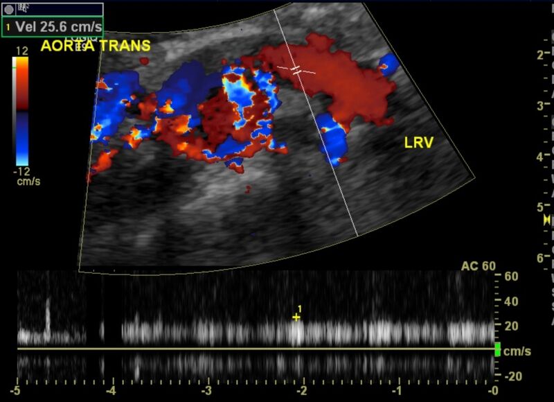
File:Nutcracker phenomenon (Radiopaedia 21159-21072 E 1).jpg
Jump to navigation
Jump to search

Size of this preview: 800 × 580 pixels. Other resolutions: 320 × 232 pixels | 640 × 464 pixels | 860 × 623 pixels.
Original file (860 × 623 pixels, file size: 125 KB, MIME type: image/jpeg)
Summary:
| Description |
|
| Date | Published: 7th Jan 2013 |
| Source | https://radiopaedia.org/cases/nutcracker-phenomenon |
| Author | Brendan Cullinane |
| Permission (Permission-reusing-text) |
http://creativecommons.org/licenses/by-nc-sa/3.0/ |
Licensing:
Attribution-NonCommercial-ShareAlike 3.0 Unported (CC BY-NC-SA 3.0)
File history
Click on a date/time to view the file as it appeared at that time.
| Date/Time | Thumbnail | Dimensions | User | Comment | |
|---|---|---|---|---|---|
| current | 17:13, 30 August 2021 | | 860 × 623 (125 KB) | Fæ (talk | contribs) | Radiopaedia project rID:21159 (thread B) (batch #26792-5 E1) |
You cannot overwrite this file.
File usage
There are no pages that use this file.
.jpg&oldid=1193976)