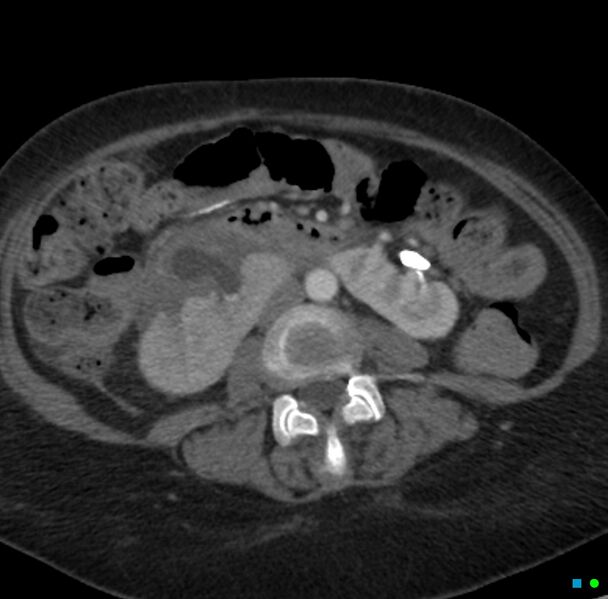
File:Obstructed infected horseshoe kidney (Radiopaedia 18116-17898 B 16).jpg
Jump to navigation
Jump to search

Size of this preview: 608 × 599 pixels. Other resolutions: 243 × 240 pixels | 487 × 480 pixels | 779 × 768 pixels | 1,186 × 1,169 pixels.
Original file (1,186 × 1,169 pixels, file size: 342 KB, MIME type: image/jpeg)
Summary:
| Description |
|
| Date | Published: 10th Jun 2012 |
| Source | https://radiopaedia.org/cases/obstructed-infected-horseshoe-kidney |
| Author | Chris O'Donnell |
| Permission (Permission-reusing-text) |
http://creativecommons.org/licenses/by-nc-sa/3.0/ |
Licensing:
Attribution-NonCommercial-ShareAlike 3.0 Unported (CC BY-NC-SA 3.0)
File history
Click on a date/time to view the file as it appeared at that time.
| Date/Time | Thumbnail | Dimensions | User | Comment | |
|---|---|---|---|---|---|
| current | 23:34, 30 August 2021 | | 1,186 × 1,169 (342 KB) | Fæ (talk | contribs) | Radiopaedia project rID:18116 (thread B) (batch #26821-42 B16) |
You cannot overwrite this file.
File usage
There are no pages that use this file.
.jpg&oldid=1196914)