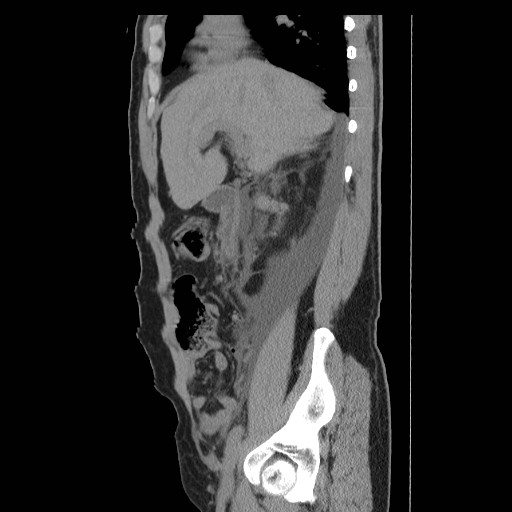
File:Obstructed kidney with perinephric urinoma (Radiopaedia 26889-27066 B 16).jpg
Jump to navigation
Jump to search
Obstructed_kidney_with_perinephric_urinoma_(Radiopaedia_26889-27066_B_16).jpg (512 × 512 pixels, file size: 42 KB, MIME type: image/jpeg)
Summary:
| Description |
|
| Date | Published: 12th Jan 2014 |
| Source | https://radiopaedia.org/cases/obstructed-kidney-with-perinephric-urinoma |
| Author | Fakhry Mahmoud Ebouda |
| Permission (Permission-reusing-text) |
http://creativecommons.org/licenses/by-nc-sa/3.0/ |
Licensing:
Attribution-NonCommercial-ShareAlike 3.0 Unported (CC BY-NC-SA 3.0)
File history
Click on a date/time to view the file as it appeared at that time.
| Date/Time | Thumbnail | Dimensions | User | Comment | |
|---|---|---|---|---|---|
| current | 23:50, 30 August 2021 |  | 512 × 512 (42 KB) | Fæ (talk | contribs) | Radiopaedia project rID:26889 (thread B) (batch #26822-80 B16) |
You cannot overwrite this file.
File usage
The following page uses this file:
