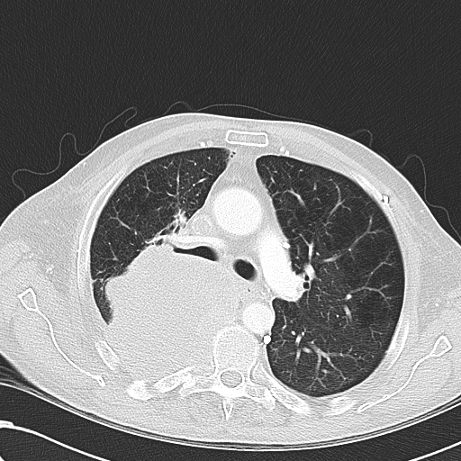
File:Obstructive superior vena cava tumor thrombus (Radiopaedia 28046-28306 Axial lung window 21).jpg
Jump to navigation
Jump to search
Obstructive_superior_vena_cava_tumor_thrombus_(Radiopaedia_28046-28306_Axial_lung_window_21).jpg (512 × 512 pixels, file size: 220 KB, MIME type: image/jpeg)
Summary:
| Description |
|
| Date | Published: 7th Mar 2014 |
| Source | https://radiopaedia.org/cases/obstructive-superior-vena-cava-tumour-thrombus |
| Author | Henry Knipe |
| Permission (Permission-reusing-text) |
http://creativecommons.org/licenses/by-nc-sa/3.0/ |
Licensing:
Attribution-NonCommercial-ShareAlike 3.0 Unported (CC BY-NC-SA 3.0)
File history
Click on a date/time to view the file as it appeared at that time.
| Date/Time | Thumbnail | Dimensions | User | Comment | |
|---|---|---|---|---|---|
| current | 23:12, 31 August 2021 |  | 512 × 512 (220 KB) | Fæ (talk | contribs) | Radiopaedia project rID:28046 (thread B) (batch #26854-127 C21) |
You cannot overwrite this file.
File usage
The following page uses this file:
