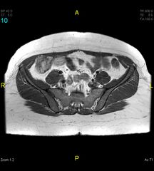
File:Obturator internus syndrome (Radiopaedia 42326-45439 Axial T1 10).jpg
Jump to navigation
Jump to search

Size of this preview: 540 × 599 pixels. Other resolutions: 216 × 240 pixels | 627 × 696 pixels.
Original file (627 × 696 pixels, file size: 60 KB, MIME type: image/jpeg)
Summary:
| Description |
|
| Date | Published: 16th Jan 2016 |
| Source | https://radiopaedia.org/cases/obturator-internus-syndrome |
| Author | Varun Babu |
| Permission (Permission-reusing-text) |
http://creativecommons.org/licenses/by-nc-sa/3.0/ |
Licensing:
Attribution-NonCommercial-ShareAlike 3.0 Unported (CC BY-NC-SA 3.0)
File history
Click on a date/time to view the file as it appeared at that time.
| Date/Time | Thumbnail | Dimensions | User | Comment | |
|---|---|---|---|---|---|
| current | 03:54, 1 September 2021 | | 627 × 696 (60 KB) | Fæ (talk | contribs) | Radiopaedia project rID:42326 (thread B) (batch #26868-10 A10) |
You cannot overwrite this file.
File usage
There are no pages that use this file.
.jpg&oldid=1218693)