File:Ollier disease - right leg (Radiopaedia 7596).jpg
Jump to navigation
Jump to search
Size of this preview: 600 × 600 pixels. Other resolutions: 240 × 240 pixels | 480 × 480 pixels | 768 × 768 pixels | 1,024 × 1,024 pixels | 1,600 × 1,600 pixels.
Original file (1,600 × 1,600 pixels, file size: 96 KB, MIME type: image/jpeg)
Summary:
- Radiopaedia case ID: 7596
- Image ID: 138185
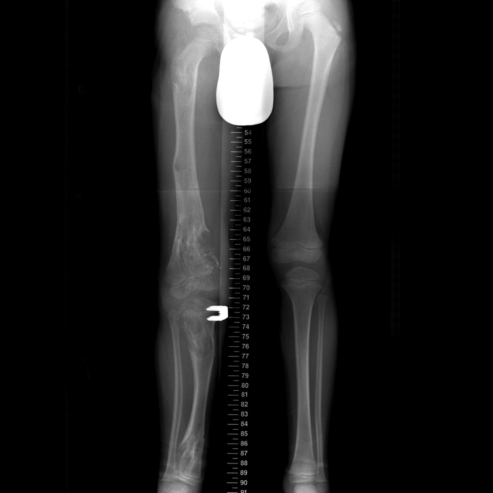
- Study findings: X-rays of the right leg of a child, demonstrates significant leg-length discrepancy with the right being shorter than the left. Involving the proximal, mid and distal femur and proximal and distal tibia are lucent lesions some of which demonstrate focal matrix mineralization. A bone staple has been inserted on the medial aspect of the tibial growth plate. Also note the unilateral involvement.
- Modality: X-ray
- System: Paediatrics
- Findings: X-rays of the right leg of a child, demonstrates significant leg-length discrepancy with the right being shorter than the left. Involving the proximal, mid and distal femur and proximal and distal tibia are lucent lesions some of which demonstrate focal matrix mineralization. A bone staple has been inserted on the medial aspect of the tibial growth plate. Also note the unilateral involvement.
- Published: 11th Nov 2009
- Source: https://radiopaedia.org/cases/ollier-disease-right-leg
- Author: Angela Byrne
- Permission: http://creativecommons.org/licenses/by-nc-sa/3.0/
Licensing:
Attribution-NonCommercial-ShareAlike 3.0 Unported (CC BY-NC-SA 3.0)
File history
Click on a date/time to view the file as it appeared at that time.
| Date/Time | Thumbnail | Dimensions | User | Comment | |
|---|---|---|---|---|---|
| current | 23:11, 24 March 2021 | | 1,600 × 1,600 (96 KB) | Fæ (talk | contribs) | Radiopaedia project rID:7596 (batch #25866) |
You cannot overwrite this file.
File usage
There are no pages that use this file.
.jpg&oldid=8855034)