File:PMC1386705 1476-7120-4-10-3.png
PMC1386705_1476-7120-4-10-3.png (512 × 248 pixels, file size: 295 KB, MIME type: image/png)
License
Attribution 2.0 Generic (CC BY 2.0)
Summary
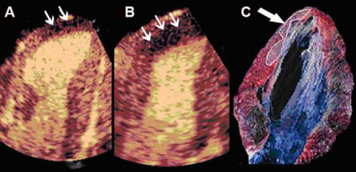
Author:Dourado PM, Tsutsui JM, Chagas AC, Sbano JC, Aiello VD, Luz PL, Mathias W, Ramires JA, Heart Institute (InCor), University of São Paulo Medical School (Openi/National library of Medicine) Source:https://openi.nlm.nih.gov/detailedresult?img=PMC1386705_1476-7120-4-10-3&query=adenosine&it=xg&req=4&npos=4 Description:F3: Representative example of real-time myocardial contrast echocardiography images showing lack of perfusion that corresponds to infarcted area before (A) and during adenosine infusion (small arrows) (B). Note that adenosine increases the infarct size determination. Necrotic area was determined as the region that failed to demonstrate brick red staining, appearing pale yellow, by triphenyl-tetrazolium chloride staining (arrow) (C).
File history
Click on a date/time to view the file as it appeared at that time.
| Date/Time | Thumbnail | Dimensions | User | Comment | |
|---|---|---|---|---|---|
| current | 23:15, 8 October 2021 | | 512 × 248 (295 KB) | Ozzie10aaaa (talk | contribs) | Author:Dourado PM, Tsutsui JM, Chagas AC, Sbano JC, Aiello VD, Luz PL, Mathias W, Ramires JA, Heart Institute (InCor), University of São Paulo Medical School (Openi/National library of Medicine) Source:https://openi.nlm.nih.gov/detailedresult?img=PMC1386705_1476-7120-4-10-3&query=adenosine&it=xg&req=4&npos=4 Description:F3: Representative example of real-time myocardial contrast echocardiography images showing lack of perfusion that corresponds to infarcted area before (A) and during adenosin... |
You cannot overwrite this file.
File usage
There are no pages that use this file.