File:PMC1523325 1750-1172-1-22-1.png
PMC1523325_1750-1172-1-22-1.png (512 × 355 pixels, file size: 330 KB, MIME type: image/png)
License
Attribution 2.0 Generic (CC BY 2.0)
Summary
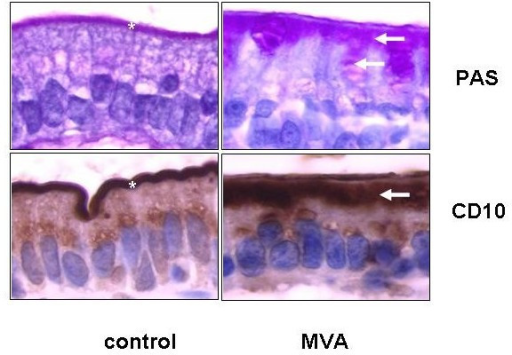
Author:Ruemmele FM, Schmitz J, Goulet O,INSERM EMI 0212, Pediatric Gastroenterology, Hepatology and Nutrition, Hôpital Necker-Enfants Malades(Openi/National Library of Medicine)Source:https://openi.nlm.nih.gov/detailedresult?img=PMC1523325_1750-1172-1-22-1&query=Microvillus%20inclusion%20disease&it=xg&req=4&npos=1 Description:F1: High power magnification of a duodenal section of a patient with typical microvillous inclusion disease or microvillous atrophy (MVA) after periodic schiff acid (PAS) staining or anti-CD10 immunohistochemistry. As shown on both panels compared to normal controls, in MVA an enlarged intracytoplasmic band (arrow) along the apical pole of enterocytes is observed along with an atrophic band instead of the normally well-defined small line representing the brush border (asterix).
File history
Click on a date/time to view the file as it appeared at that time.
| Date/Time | Thumbnail | Dimensions | User | Comment | |
|---|---|---|---|---|---|
| current | 21:55, 23 January 2022 | | 512 × 355 (330 KB) | Ozzie10aaaa (talk | contribs) | Author:Ruemmele FM, Schmitz J, Goulet O,INSERM EMI 0212, Pediatric Gastroenterology, Hepatology and Nutrition, Hôpital Necker-Enfants Malades(Openi/National Library of Medicine)Source:https://openi.nlm.nih.gov/detailedresult?img=PMC1523325_1750-1172-1-22-1&query=Microvillus%20inclusion%20disease&it=xg&req=4&npos=1 Description:F1: High power magnification of a duodenal section of a patient with typical microvillous inclusion disease or microvillous atrophy (MVA) after periodic schiff acid (PAS... |
You cannot overwrite this file.
File usage
There are no pages that use this file.