File:PMC1808442 1750-1172-2-7-3.png
PMC1808442_1750-1172-2-7-3.png (512 × 425 pixels, file size: 481 KB, MIME type: image/png)
License
Attribution 2.0 Generic (CC BY 2.0)
Summary
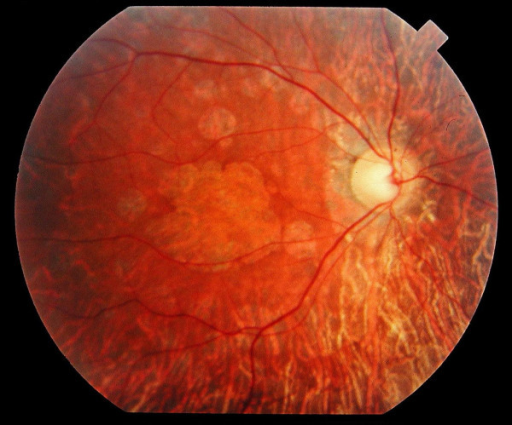
Author:Hamel CP, Inserm U. 583, Physiopathologie et thérapie des déficits sensoriels et moteurs, Institut des Neurosciences de Montpellier(Openi/National Library of Medicine) Source: https://openi.nlm.nih.gov/detailedresult?img=PMC1808442_1750-1172-2-7-3&query=Spinocerebellar%20ataxia&it=xg&req=4&npos=6 Description:F3: Fundus of a 34 year-old patient with cone rod dystrophy due to Spinocerebellar Ataxia Type 7 (SCA7). Note that the macular area, and also the mid periphery, are atrophic.
File history
Click on a date/time to view the file as it appeared at that time.
| Date/Time | Thumbnail | Dimensions | User | Comment | |
|---|---|---|---|---|---|
| current | 23:47, 5 February 2022 | | 512 × 425 (481 KB) | Ozzie10aaaa (talk | contribs) | Author:Hamel CP, Inserm U. 583, Physiopathologie et thérapie des déficits sensoriels et moteurs, Institut des Neurosciences de Montpellier(Openi/National Library of Medicine) Source: https://openi.nlm.nih.gov/detailedresult?img=PMC1808442_1750-1172-2-7-3&query=Spinocerebellar%20ataxia&it=xg&req=4&npos=6 Description:F3: Fundus of a 34 year-old patient with cone rod dystrophy due to Spinocerebellar Ataxia Type 7 (SCA7). Note that the macular area, and also the mid periphery, are atrophic. |
You cannot overwrite this file.
File usage
There are no pages that use this file.