File:PMC2519249 ehn29605.png
PMC2519249_ehn29605.png (400 × 251 pixels, file size: 80 KB, MIME type: image/png)
License
Attribution 4.0 International (CC BY 4.0)
Summary
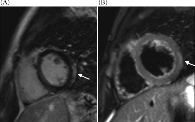
Author:Dennert R, Crijns HJ, Heymans S, Department of Cardiology, CARIM, University Hospital(Openi/National Library of Medicine) Source:https://openi.nlm.nih.gov/detailedresult?img=PMC2519249_ehn29605&query=Autoimmune%20myocarditis&it=xg&req=4&npos=70 Description:EHN296F5: Cardiovascular magnetic resonance image. Short-axis cardiac magnetic resonance imaging of a patient with acute myocarditis (A) T2-weighted image, showing regional oedema of the lateral left ventricle predominantly subepicardial involvement (arrow). (B) Late enhancement image, demonstrating high signal intensity in the epicardial region of the lateral wall of the left ventricle (arrow).
File history
Click on a date/time to view the file as it appeared at that time.
| Date/Time | Thumbnail | Dimensions | User | Comment | |
|---|---|---|---|---|---|
| current | 20:51, 2 January 2022 | | 400 × 251 (80 KB) | Ozzie10aaaa (talk | contribs) | Author:Dennert R, Crijns HJ, Heymans S, Department of Cardiology, CARIM, University Hospital(Openi/National Library of Medicine) Source:https://openi.nlm.nih.gov/detailedresult?img=PMC2519249_ehn29605&query=Autoimmune%20myocarditis&it=xg&req=4&npos=70 Description:EHN296F5: Cardiovascular magnetic resonance image. Short-axis cardiac magnetic resonance imaging of a patient with acute myocarditis (A) T2-weighted image, showing regional oedema of the lateral left ventricle predominantly subepicar... |
You cannot overwrite this file.
File usage
There are no pages that use this file.