File:PMC2627172 kjr-9-91-g001 (1).png
PMC2627172_kjr-9-91-g001_(1).png (497 × 548 pixels, file size: 122 KB, MIME type: image/png)
License
Attribution-NonCommercial 3.0 Unported (CC BY-NC 3.0)
Summary
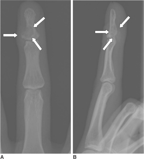
Author:Choi SJ, Ahn JH, Kang G, Lee JH, Park MS, Ryu DS, Jung SM ,Department of Radiology, Asan Foundation, GangNeung Asan Hospital, University of Ulsan College of Medicine(Openi/National Library of Medicine)Source:https://openi.nlm.nih.gov/detailedresult?img=PMC2627172_kjr-9-91-g001&query=Aponeurotic%20fibroma&it=xg&req=4&npos=3 Description:F1: Left middle finger AP (A) and lateral (B) views show eccentrically located well-defined osteolytic lesion in the base of the distal phalanx (arrows). Calcific foci are noted in the mass and soft tissue mass component is obvious. On these radiographs, soft tissue mass with large cortical erosion is indistinguishable with eccentrically locating osteolytic mass with soft tissue extension.
File history
Click on a date/time to view the file as it appeared at that time.
| Date/Time | Thumbnail | Dimensions | User | Comment | |
|---|---|---|---|---|---|
| current | 19:40, 21 April 2022 | | 497 × 548 (122 KB) | Ozzie10aaaa (talk | contribs) | Author:Choi SJ, Ahn JH, Kang G, Lee JH, Park MS, Ryu DS, Jung SM ,Department of Radiology, Asan Foundation, GangNeung Asan Hospital, University of Ulsan College of Medicine(Openi/National Library of Medicine)Source:https://openi.nlm.nih.gov/detailedresult?img=PMC2627172_kjr-9-91-g001&query=Aponeurotic%20fibroma&it=xg&req=4&npos=3 Description:F1: Left middle finger AP (A) and lateral (B) views show eccentrically located well-defined osteolytic lesion in the base of the distal phalanx (arrows).... |
You cannot overwrite this file.
File usage
There are no pages that use this file.
.png&oldid=1247401)