File:PMC2713834 kjr-3-113-g003.png
PMC2713834_kjr-3-113-g003.png (512 × 444 pixels, file size: 156 KB, MIME type: image/png)
License
Attribution-NonCommercial 3.0 Unported (CC BY-NC 3.0)
Summary
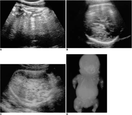
Author:Lee SH, Cho JY, Song MJ, Min JY, Han BH, Lee YH, Cho BJ, Kim SH, Department of Radiology, Samsung Cheil Hospital, Sungkyunkwan University School of Medicine (Openi/National Library of medicine) Source:https://openi.nlm.nih.gov/detailedresult?img=PMC2713834_kjr-3-113-g003&query=Achondrogenesis%20type%202&it=xg&req=4&npos=6 Description:F3: Achondrogenesis in a 35-week fetus.A. Ultrasonogram shows profound limb shortening (arrow).B, C. Axial images of the fetal head demonstrate decreased calvarial ossification, with increased ultrasonic through-transmission. The fetal head is compressed by the transducer.D. Postmortem radiograph shows extremely short limbs, a large head, and the absence of ossification in the ischia, pubis, vertebral body, and calvarium. The head is disproportionately enlarged and the thorax is small.
File history
Click on a date/time to view the file as it appeared at that time.
| Date/Time | Thumbnail | Dimensions | User | Comment | |
|---|---|---|---|---|---|
| current | 00:25, 9 February 2022 | | 512 × 444 (156 KB) | Ozzie10aaaa (talk | contribs) | Author:Lee SH, Cho JY, Song MJ, Min JY, Han BH, Lee YH, Cho BJ, Kim SH, Department of Radiology, Samsung Cheil Hospital, Sungkyunkwan University School of Medicine (Openi/National Library of medicine) Source:https://openi.nlm.nih.gov/detailedresult?img=PMC2713834_kjr-3-113-g003&query=Achondrogenesis%20type%202&it=xg&req=4&npos=6 Description:F3: Achondrogenesis in a 35-week fetus.A. Ultrasonogram shows profound limb shortening (arrow).B, C. Axial images of the fetal head demonstrate decreased... |
You cannot overwrite this file.
File usage
There are no pages that use this file.