File:PMC2823763 1752-1947-4-8-2.png
PMC2823763_1752-1947-4-8-2.png (512 × 197 pixels, file size: 252 KB, MIME type: image/png)
License
Attribution 2.0 Generic (CC BY 2.0)
Summary
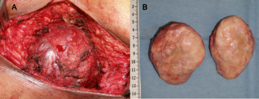
Author:Kitahata Y, Yokoyama S, Takifuji K, Hotta T, Matsuda K, Tominaga T, Oku Y, Watanabe T, Ieda J, Yamaue H,Second Department of Surgery, Wakayama Medical University, School of Medicine (Openi/National Library of Medicine) Source:https://openi.nlm.nih.gov/detailedresult?img=PMC2823763_1752-1947-4-8-2&query=Hemangiopericytoma&it=xg&req=4&npos=55 Description:F2: (A) This is a macroscopic image of the 80 × 75 × 65 mm-sized tumor in the sacrococcygeal space. (B) This image shows the excised tumor with a capsule, with its cut surface mostly grayish white and partially reddish.
File history
Click on a date/time to view the file as it appeared at that time.
| Date/Time | Thumbnail | Dimensions | User | Comment | |
|---|---|---|---|---|---|
| current | 22:09, 24 September 2021 | 512 × 197 (252 KB) | Ozzie10aaaa (talk | contribs) | Author:Kitahata Y, Yokoyama S, Takifuji K, Hotta T, Matsuda K, Tominaga T, Oku Y, Watanabe T, Ieda J, Yamaue H,Second Department of Surgery, Wakayama Medical University, School of Medicine (Openi/National Library of Medicine) Source:https://openi.nlm.nih.gov/detailedresult?img=PMC2823763_1752-1947-4-8-2&query=Hemangiopericytoma&it=xg&req=4&npos=55 Description:F2: (A) This is a macroscopic image of the 80 × 75 × 65 mm-sized tumor in the sacrococcygeal space. (B) This image shows the excised tu... |
You cannot overwrite this file.
File usage
There are no pages that use this file.