File:PMC2828421 1471-2474-11-18-2.png
PMC2828421_1471-2474-11-18-2.png (512 × 196 pixels, file size: 190 KB, MIME type: image/png)
License
Attribution 2.0 Generic (CC BY 2.0)
Summary
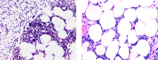
Author:Pongratz G, Ehrenstein B, Hartung W, Schölmerich J, Fleck M,Department of Internal Medicine I, University Medical Center Regensburg (Openi/National Library of Medicine) Source:https://openi.nlm.nih.gov/detailedresult?img=PMC2828421_1471-2474-11-18-2&query=Weber%E2%80%93Christian%20disease&it=xg&req=4&npos=1 Description:F2: Histology (painful node, left lower leg). Standard histological processing was performed on a painful node obtained from the lower limb. Two HE-stained sections at different magnifications (left 20×, right 30×) are depicted. The sections show predominantly lobular panniculitis with mixed inflammatory cell infiltration.
File history
Click on a date/time to view the file as it appeared at that time.
| Date/Time | Thumbnail | Dimensions | User | Comment | |
|---|---|---|---|---|---|
| current | 22:22, 1 March 2022 | 512 × 196 (190 KB) | Ozzie10aaaa (talk | contribs) | Author:Pongratz G, Ehrenstein B, Hartung W, Schölmerich J, Fleck M,Department of Internal Medicine I, University Medical Center Regensburg (Openi/National Library of Medicine) Source:https://openi.nlm.nih.gov/detailedresult?img=PMC2828421_1471-2474-11-18-2&query=Weber%E2%80%93Christian%20disease&it=xg&req=4&npos=1 Description:F2: Histology (painful node, left lower leg). Standard histological processing was performed on a painful node obtained from the lower limb. Two HE-stained sections at d... |
You cannot overwrite this file.
File usage
There are no pages that use this file.