File:PMC2958498 IJHT2010-964513.002.png
PMC2958498_IJHT2010-964513.002.png (478 × 339 pixels, file size: 190 KB, MIME type: image/png)
License
Attribution-NonCommercial 4.0 International (CC BY-NC 4.0)
Summary
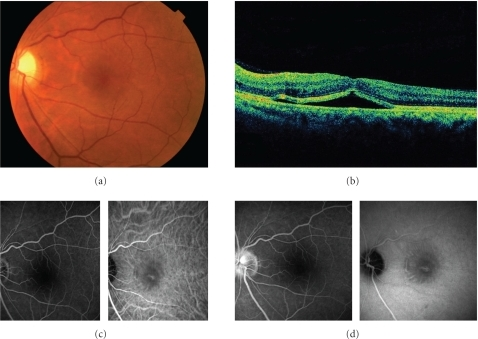
Author:Hirano Y, Yasukawa T, Ogura Y, Department of Ophthalmology & Visual Science, Nagoya City University Graduate School of Medical Sciences (Openi/National Library of medicine) Source:https://openi.nlm.nih.gov/detailedresult?img=PMC2958498_IJHT2010-964513.002&query=retinal%20detachment&it=xg&req=4&npos=4 Description:fig2: The left eye of the same patient at the first visit. (a) Fundus photograph shows serous retinal detachment. The optic disc is swelling. (b) OCT shows retinal detachment involving the fovea and cystic change of inner retina. (c) Early phase images of FA (left) and IA (right). (d) Late phase images of FA (left) and IA (right). Note that FA shows no active leakage corresponding to the area with serous retinal detachment and staining of the swollen optic disc. IA shows decreased perfusion of the choroid at the macula and window defect associated with damaged RPE.
File history
Click on a date/time to view the file as it appeared at that time.
| Date/Time | Thumbnail | Dimensions | User | Comment | |
|---|---|---|---|---|---|
| current | 00:45, 26 February 2022 | | 478 × 339 (190 KB) | Ozzie10aaaa (talk | contribs) | Author:Hirano Y, Yasukawa T, Ogura Y, Department of Ophthalmology & Visual Science, Nagoya City University Graduate School of Medical Sciences (Openi/National Library of medicine) Source:https://openi.nlm.nih.gov/detailedresult?img=PMC2958498_IJHT2010-964513.002&query=retinal%20detachment&it=xg&req=4&npos=4 Description:fig2: The left eye of the same patient at the first visit. (a) Fundus photograph shows serous retinal detachment. The optic disc is swelling. (b) OCT shows retinal detachment i... |
You cannot overwrite this file.
File usage
There are no pages that use this file.