File:PMC2998243 cia-5-365f1.png
PMC2998243_cia-5-365f1.png (395 × 403 pixels, file size: 135 KB, MIME type: image/png)
License
Attribution-NonCommercial 3.0 Unported (CC BY-NC 3.0)
Summary
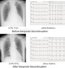
Author:Yanagisawa S, Suzuki N, Tanaka T,Department of Cardiology, Okazaki City Hospital(Openi/National Library of Medicine) Source:https://openi.nlm.nih.gov/detailedresult?img=PMC2998243_cia-5-365f1&query=Bisoprolol&it=xg&req=4&npos=4 Description:f1-cia-5-365: At the time of the most severe condition, the chest radiograph showed cardiomegaly with cardiothoracic ratio (CTR) of 71%; an electrocardiogram revealed atrial fibrillation with a QS pattern in the V1–V3 leads A). After bisoprolol discontinuation, the CTR determined by chest radiography was reduced to 57%, and atrial fibrillation converted to sinus rhythm B).
{{{category}}}
File history
Click on a date/time to view the file as it appeared at that time.
| Date/Time | Thumbnail | Dimensions | User | Comment | |
|---|---|---|---|---|---|
| current | 22:32, 21 January 2022 |  | 395 × 403 (135 KB) | Ozzie10aaaa (talk | contribs) | Author:Yanagisawa S, Suzuki N, Tanaka T,Department of Cardiology, Okazaki City Hospital(Openi/National Library of Medicine) Source:https://openi.nlm.nih.gov/detailedresult?img=PMC2998243_cia-5-365f1&query=Bisoprolol&it=xg&req=4&npos=4 Description:f1-cia-5-365: At the time of the most severe condition, the chest radiograph showed cardiomegaly with cardiothoracic ratio (CTR) of 71%; an electrocardiogram revealed atrial fibrillation with a QS pattern in the V1–V3 leads A). After bisoprolol disco... |
You cannot overwrite this file.
File usage
The following page uses this file:

