File:PMC3016775 03-0367-F1 (1).png
Jump to navigation
Jump to search
No higher resolution available.
PMC3016775_03-0367-F1_(1).png (238 × 173 pixels, file size: 24 KB, MIME type: image/png)
Summary
| Description |
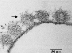
English: F1: Thin-section electron micrograph of severe acute respiratory syndrome-associated coronavirus grown in Vero E6 cells. Panel A shows extracellular viral particles (arrow) lining the surface of the plasma membrane. Some spikes projecting from the envelope of the virus are seen. Panel B shows numerous spherical coronavirus particles (arrow) within dilated cytoplasmic vacuoles (arrowhead). |
| Date | |
| Source | https://openi.nlm.nih.gov/detailedresult?img=PMC3016775_03-0367-F1&query=coronavirus&it=xg&req=4&npos=31 |
| Author | Hsueh PR, Hsiao CH, Yeh SH, Wang WK, Chen PJ, Wang JT, Chang SC, Kao CL, Yang PC, SARS Research Group of National Taiwan University College of Medicine and National Taiwan University Hospit |
Licensing
{{subst:Custom license marker added by UW}} https://creativecommons.org/publicdomain/zero/1.0/ CC0 1.0 Universal (CC0 1.0) Public Domain Dedication
This file was uploaded with UploadWizard.
File history
Click on a date/time to view the file as it appeared at that time.
| Date/Time | Thumbnail | Dimensions | User | Comment | |
|---|---|---|---|---|---|
| current | 20:38, 15 March 2023 | | 238 × 173 (24 KB) | Ozzie10aaaa (talk | contribs) | Uploaded a work by Hsueh PR, Hsiao CH, Yeh SH, Wang WK, Chen PJ, Wang JT, Chang SC, Kao CL, Yang PC, SARS Research Group of National Taiwan University College of Medicine and National Taiwan University Hospit from https://openi.nlm.nih.gov/detailedresult?img=PMC3016775_03-0367-F1&query=coronavirus&it=xg&req=4&npos=31 with UploadWizard |
You cannot overwrite this file.
File usage
There are no pages that use this file.
.png&oldid=1252130)