File:PMC3034459 ipej100556-01.png
Jump to navigation
Jump to search
No higher resolution available.
PMC3034459_ipej100556-01.png (512 × 275 pixels, file size: 160 KB, MIME type: image/png)
Summary
| Description |
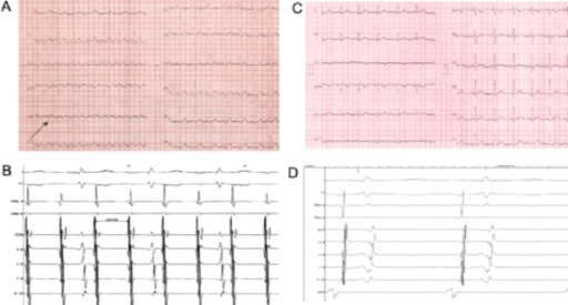
English: F1: Basal ECG (A): atrial tachycardia with 2:1 conduction is detectable. The atrial waves morphology (negative in lateral leads - see black arrow - and positive in V1) involves the likely diagnosis of left sided atrial tachycardia. Intracardiac electrograms (B) show left atrial tachycardia (cycle length 300 msec) with 2:1 conduction to the ventricles; the distal dipole of the catheter positioned inside the coronary sinus is the first one to be recording atrial signals, thus suggesting a left lateral origin of the tachycardia. The registration speed is 100 mm/sec. Post-procedural surface ECG (C): sinus rhythm is visible. Intracardiac electrograms (D) confirm the earliest atrial activation to be at the level of the high right atrium (HRA). Map d = distal dipole of the ablation catheter. Map p = proximal dipole of the ablation catheter. DCS = distal dipole of the catheter in coronary sinus. HRA = catheter positioned at the level of the high right atrium |
| Date | |
| Source | https://openi.nlm.nih.gov/detailedresult?img=PMC3034459_ipej100556-01&query=Atrial%20tachycardia&it=xg&req=4&npos=2 |
| Author | Pandozi C, Galeazzi M, Lavalle C, Ficili S, Russo M, Santini M |
Licensing
English: This file is licensed CC BY-NC 4.0
This file was uploaded with UploadWizard.
File history
Click on a date/time to view the file as it appeared at that time.
| Date/Time | Thumbnail | Dimensions | User | Comment | |
|---|---|---|---|---|---|
| current | 20:04, 23 October 2022 | | 512 × 275 (160 KB) | Ozzie10aaaa (talk | contribs) | Uploaded a work by Pandozi C, Galeazzi M, Lavalle C, Ficili S, Russo M, Santini M from https://openi.nlm.nih.gov/detailedresult?img=PMC3034459_ipej100556-01&query=Atrial%20tachycardia&it=xg&req=4&npos=2 with UploadWizard |
You cannot overwrite this file.
File usage
There are no pages that use this file.