File:PMC3068520 415 2010 5775 Fig1 HTML.png
Jump to navigation
Jump to search
No higher resolution available.
PMC3068520_415_2010_5775_Fig1_HTML.png (480 × 397 pixels, file size: 196 KB, MIME type: image/png)
Summary
| Description |
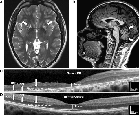
English: Fig1: High-resolution retinal and brain imaging in NARP syndrome demonstrates analogous patterns of tissue injury. This 28-year-old woman (D1) with NARP syndrome from the mtDNA ATPase 6 m.8993T>C mutation with 78% blood leukocyte and 99% hair-bulb heteroplasmy had severe RP and moderate ataxia. a, b 3-T MRI demonstrates cystic and cavitary T2 hyperintensities in the bilateral putamina (a), likely reflecting neuronal necrosis, and also moderate cerebellar atrophy with T1 imaging (b). c High-resolution OCT image of the macula demonstrates severe retinal thinning, primarily due to degeneration of the photoreceptor and the retinal pigment epithelial cell layers, but also associated thinning of the ganglion cell (GCL) and retinal nerve fiber layer (RNFL). d Macular OCT image from an age-similar normal female shown for comparison |
| Date | |
| Source | https://openi.nlm.nih.gov/detailedresult?img=PMC3068520_415_2010_5775_Fig1_HTML&query=NARP%20syndrome&it=xg&req=4&npos=2 |
| Author | Gelfand JM, Duncan JL, Racine CA, Gillum LA, Chin CT, Zhang Y, Zhang Q, Wong LJ, Roorda A, Green AJ |
Licensing
Creative Commons Attribution-NonCommercial-ShareAlike 4.0 International
This file was uploaded with UploadWizard.
File history
Click on a date/time to view the file as it appeared at that time.
| Date/Time | Thumbnail | Dimensions | User | Comment | |
|---|---|---|---|---|---|
| current | 18:45, 6 September 2022 | | 480 × 397 (196 KB) | Ozzie10aaaa (talk | contribs) | Uploaded a work by Gelfand JM, Duncan JL, Racine CA, Gillum LA, Chin CT, Zhang Y, Zhang Q, Wong LJ, Roorda A, Green AJ from https://openi.nlm.nih.gov/detailedresult?img=PMC3068520_415_2010_5775_Fig1_HTML&query=NARP%20syndrome&it=xg&req=4&npos=2 with UploadWizard |
You cannot overwrite this file.
File usage
There are no pages that use this file.