File:PMC3068591 JGID-3-96-g001.png
Jump to navigation
Jump to search

Size of this preview: 270 × 598 pixels. Other resolutions: 108 × 240 pixels | 462 × 1,024 pixels.
Original file (462 × 1,024 pixels, file size: 303 KB, MIME type: image/png)
Summary
| Description |
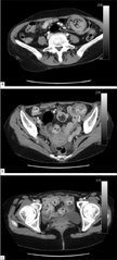
English: F1: (a-c) Axial CT scans of the abdomen showing multiple intussusceptions (asterisks) in the jejunum and ileum caused by Kaposi's sarcoma; (a) Image shows round mass with a target pattern (“bowel-within-bowel appearance”) due to adjacent bowel walls of the outer intussuscipiens (double arrows) and the inner intussusceptum (arrow). Inside, a half-moon-shaped hypodense area of fat density representing mesenteric fat, plus vessels (solid arrowhead). This pattern is observed when axis of intussusception is perpendicular to CT beam. (b, c) Images show several enteroenteric intussusception forming several loops with the abdomen and pelvis. In some, a lead point corresponding to Kaposi's submucosal masses is seen (open arrowhead) |
| Date | |
| Source | https://openi.nlm.nih.gov/detailedresult?img=PMC3068591_JGID-3-96-g001&query=Intussusception&it=xg&req=4&npos=1 |
| Author | Afonso PD, Lourenço R |
Licensing
English: This file is licensed CC BY-NC-SA 3.0
This file was uploaded with UploadWizard.
File history
Click on a date/time to view the file as it appeared at that time.
| Date/Time | Thumbnail | Dimensions | User | Comment | |
|---|---|---|---|---|---|
| current | 21:28, 27 December 2022 | | 462 × 1,024 (303 KB) | Ozzie10aaaa (talk | contribs) | Uploaded a work by Afonso PD, Lourenço R from https://openi.nlm.nih.gov/detailedresult?img=PMC3068591_JGID-3-96-g001&query=Intussusception&it=xg&req=4&npos=1 with UploadWizard |
You cannot overwrite this file.
File usage
There are no pages that use this file.