File:PMC3078321 431 2011 1452 Fig1 HTML.png
PMC3078321_431_2011_1452_Fig1_HTML.png (496 × 372 pixels, file size: 256 KB, MIME type: image/png)
License
Attribution-NonCommercial 3.0 Unported (CC BY-NC 3.0)
Summary
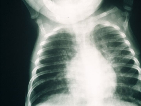
Author:van der Burg M, Gennery AR, Department of Immunology, Erasmus MC, University Medical Center Rotterdam(Openi/National Library of Medicine) Source:https://openi.nlm.nih.gov/detailedresult?img=PMC3078321_431_2011_1452_Fig1_HTML&query=Severe%20combined%20immunodeficiency&it=xg&req=4&npos=1 Description:Fig1: Chest radiograph from a 5-month-old infant with severe combined immunodeficiency showing bilateral patchy shadowing secondary to interstitial pnuemonitis due to infection with respiratory syncytial virus and Pneumocystis jiroveci. There is hyperinflation of the lungs, and the midline pleural borders of the upper lobes are visible because the thymic shadow is absent (courtesy of The Paediatric Immunology Unit, Great North Children’s Hospital, Newcastle upon Tyne)
File history
Click on a date/time to view the file as it appeared at that time.
| Date/Time | Thumbnail | Dimensions | User | Comment | |
|---|---|---|---|---|---|
| current | 23:16, 6 February 2022 | | 496 × 372 (256 KB) | Ozzie10aaaa (talk | contribs) | Author:van der Burg M, Gennery AR, Department of Immunology, Erasmus MC, University Medical Center Rotterdam(Openi/National Library of Medicine) Source:https://openi.nlm.nih.gov/detailedresult?img=PMC3078321_431_2011_1452_Fig1_HTML&query=Severe%20combined%20immunodeficiency&it=xg&req=4&npos=1 Description:Fig1: Chest radiograph from a 5-month-old infant with severe combined immunodeficiency showing bilateral patchy shadowing secondary to interstitial pnuemonitis due to infection with respirato... |
You cannot overwrite this file.
File usage
There are no pages that use this file.