File:PMC3153151 tmh-39-1-suppl 2-65-g002.png
Jump to navigation
Jump to search
No higher resolution available.
PMC3153151_tmh-39-1-suppl_2-65-g002.png (512 × 286 pixels, file size: 106 KB, MIME type: image/png)
Summary
| Description |
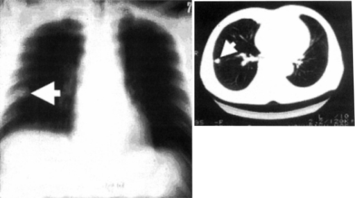
English: F2: Chest X-ray (left) and CT (right) appearance of a patient with pulmonary dirofilariasis. A solitary nodule called “coin lesion” is adjacent to the pleural membrane (arrow). |
| Date | |
| Source | https://openi.nlm.nih.gov/detailedresult?img=PMC3153151_tmh-39-1-suppl_2-65-g002&query=Dirofilariasis&it=xg&req=4&npos=2 |
| Author | Akao N |
Licensing
English: This file is licensed CC BY-NC 3.0
This file was uploaded with UploadWizard.
File history
Click on a date/time to view the file as it appeared at that time.
| Date/Time | Thumbnail | Dimensions | User | Comment | |
|---|---|---|---|---|---|
| current | 20:09, 30 January 2023 | | 512 × 286 (106 KB) | Ozzie10aaaa (talk | contribs) | Uploaded a work by Akao N from https://openi.nlm.nih.gov/detailedresult?img=PMC3153151_tmh-39-1-suppl_2-65-g002&query=Dirofilariasis&it=xg&req=4&npos=2 with UploadWizard |
You cannot overwrite this file.
File usage
There are no pages that use this file.