File:PMC3168806 kjr-12-629-g001 (1) (1) (1).png
Jump to navigation
Jump to search
No higher resolution available.
PMC3168806_kjr-12-629-g001_(1)_(1)_(1).png (250 × 350 pixels, file size: 29 KB, MIME type: image/png)
Summary
| Description |
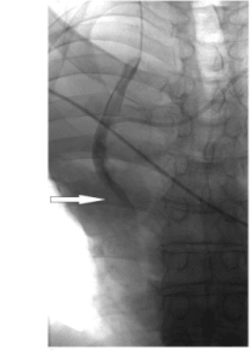
English: F1: Angiographic and schematic illustration of procedure.A. Diagnostic angiography showing severe stenosis of superior vena cava (arrow). B. Diagnostic angiography of left brachiocephalic vein reveals subtotal stenosis (arrows). C. After repositioning tip of port-catheter into right brachiocephalic vein using 15 mm Andra-snare, self-expandable stent is placed in superior vena cava (arrows). D. Left jugular vein is punctured and 7 Fr introducer sheath is introduced (arrowhead). Released stent in superior vena cava (arrow). E. Using Andra-snare (arrow), port-catheter (arrowhead) is repositioned in distal part of left brachiocephalic vein and is fixed there by keeping loop-wire of snare around port catheter, while snare's catheter is removed. F. Left brachiocephalic vein stenosis is crossed with guide-wire via same sheath while snare's loop wire remains in place and two 12 mm stents (arrow) are placed via same introducer sheath in left brachiocephalic vein, extending from superior vena cava to left brachiocephalic vein. Finally snare's catheter is inserted again and port catheter is repositioned into superior vena cava (arrowhead). G-J. Schematic drawing of procedure. Initial situation with stenosis in superior vena cava and left brachiocephalic vein and in situ port catheter (G). Port catheter is repositioned in right brachiocephalic vein using snare before stent is placed in superior vena cava (H). Then port catheter is positioned in distal part of left brachiocephalic vein and two stents are placed in left brachiocephalic vein and superior vena cava via same sheath (I). Final situation with preserved port catheter (J). |
| Date | |
| Source | https://openi.nlm.nih.gov/detailedresult?img=PMC3168806_kjr-12-629-g001&query=stenosis%20of%20vena%20cava&it=xg&req=4&npos=1 |
| Author | Isfort P, Penzkofer T, Goerg F, Mahnken AH |
Licensing
English: This file is licensed CC BY-NC 3.0
This file was uploaded with UploadWizard.
File history
Click on a date/time to view the file as it appeared at that time.
| Date/Time | Thumbnail | Dimensions | User | Comment | |
|---|---|---|---|---|---|
| current | 17:34, 1 December 2022 | | 250 × 350 (29 KB) | Ozzie10aaaa (talk | contribs) | Uploaded a work by Isfort P, Penzkofer T, Goerg F, Mahnken AH from https://openi.nlm.nih.gov/detailedresult?img=PMC3168806_kjr-12-629-g001&query=stenosis%20of%20vena%20cava&it=xg&req=4&npos=1 with UploadWizard |
You cannot overwrite this file.
File usage
There are no pages that use this file.
_(1)_(1).png&oldid=1251261)