File:PMC3177427 JCIS-1-20-g002.png
PMC3177427_JCIS-1-20-g002.png (512 × 177 pixels, file size: 85 KB, MIME type: image/png)
License
Attribution-NonCommercial-ShareAlike 3.0 Unported (CC BY-NC-SA 3.0)
Summary
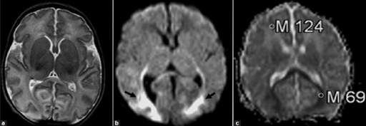
Author:Sener RN, Atalar MH,Department of Radiology, Ege University School of Medicine (Openi/National Library of medicine) Source:https://openi.nlm.nih.gov/detailedresult?img=PMC3177427_JCIS-1-20-g002&query=Neonatal%20adrenoleukodystrophy&it=xg&req=4&npos=1 Description:F1: Neonatal adrenoleukodystrophy: (a) the axial T2-weighted MR image is normal; (b) the diffusion-weighted image (b = 1000 sec/mm2) reveals high-signal changes in the splenium of the corpus callosum, and occipital lobes (black arrows); (c) the ADC map reveals that the ADC values of the involved regions is low (0.69 × 10⁻³ mm2/sec), compared to the frontal regions (1.24 × 10⁻³ mm2/s).
File history
Click on a date/time to view the file as it appeared at that time.
| Date/Time | Thumbnail | Dimensions | User | Comment | |
|---|---|---|---|---|---|
| current | 19:21, 27 August 2021 | 512 × 177 (85 KB) | Ozzie10aaaa (talk | contribs) | Author:Sener RN, Atalar MH,Department of Radiology, Ege University School of Medicine (Openi/National Library of medicine) Source:https://openi.nlm.nih.gov/detailedresult?img=PMC3177427_JCIS-1-20-g002&query=Neonatal%20adrenoleukodystrophy&it=xg&req=4&npos=1 Description:F1: Neonatal adrenoleukodystrophy: (a) the axial T2-weighted MR image is normal; (b) the diffusion-weighted image (b = 1000 sec/mm2) reveals high-signal changes in the splenium of the corpus callosum, and occipital lobes (black... |
You cannot overwrite this file.
File usage
There are no pages that use this file.