File:PMC3212896 1750-1172-6-68-1.png
Jump to navigation
Jump to search
No higher resolution available.
PMC3212896_1750-1172-6-68-1.png (512 × 407 pixels, file size: 317 KB, MIME type: image/png)
License
Attribution 2.0 Generic (CC BY 2.0)
Summary
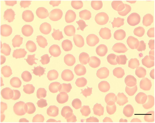
Author:Jung HH, Danek A, Walker RH, Department of Neurology, University Hospital Zürich(Openi/National Library of Medicine) Source:https://openi.nlm.nih.gov/detailedresult?img=PMC3212896_1750-1172-6-68-1&query=McLeod%20syndrome&it=xg&req=4&npos=1 Description:F1: Acanthocytes. Peripheral blood smear showing acanthocytosis in a patient with McLeod syndrome (May Gruenwald-Giemsa; x100; scale bar = 10 μm).
File history
Click on a date/time to view the file as it appeared at that time.
| Date/Time | Thumbnail | Dimensions | User | Comment | |
|---|---|---|---|---|---|
| current | 23:41, 10 February 2022 | | 512 × 407 (317 KB) | Ozzie10aaaa (talk | contribs) | Author:Jung HH, Danek A, Walker RH, Department of Neurology, University Hospital Zürich(Openi/National Library of Medicine) Source:https://openi.nlm.nih.gov/detailedresult?img=PMC3212896_1750-1172-6-68-1&query=McLeod%20syndrome&it=xg&req=4&npos=1 Description:F1: Acanthocytes. Peripheral blood smear showing acanthocytosis in a patient with McLeod syndrome (May Gruenwald-Giemsa; x100; scale bar = 10 μm). |
You cannot overwrite this file.
File usage
There are no pages that use this file.