File:PMC3257870 fnhum-05-00138-g001.png
Jump to navigation
Jump to search
No higher resolution available.
PMC3257870_fnhum-05-00138-g001.png (491 × 179 pixels, file size: 55 KB, MIME type: image/png)
Summary
| Description |
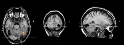
English: F1: Brain damage profile of the acquired prosopagnosia patient PS. This patient has damage to the left middle fusiform gyrus and a small lesion to the right middle temporal gyrus, but her main lesion, thought to be instrumental in causing her prosopagnosia, concerns the right inferior occipital cortex (line crossing). This lesion does not prevent a preferential activation for faces in the right middle fusiform gyrus (FFA; here as the result of a combined analysis of six functional localizer runs, contrasting faces and object pictures in a face localizer contrast, see Sorger et al., 2007 for details). Although this region responds preferentially to faces, it does not show the normal release to adaptation to different facial identities, in line with the difficulties of the patient in individualizing faces. TRA, transverse plane; COR, coronal plane; SAG, sagittal plane; R, right. |
| Date | |
| Source | https://openi.nlm.nih.gov/detailedresult?img=PMC3257870_fnhum-05-00138-g001&query=Prosopagnosia&it=xg&req=4&npos=1 |
| Author | Prieto EA, Caharel S, Henson R, Rossion B |
Licensing
English: This file is licensed CC BY-NC 4.0
This file was uploaded with UploadWizard.
File history
Click on a date/time to view the file as it appeared at that time.
| Date/Time | Thumbnail | Dimensions | User | Comment | |
|---|---|---|---|---|---|
| current | 21:55, 15 February 2023 | 491 × 179 (55 KB) | Ozzie10aaaa (talk | contribs) | Uploaded a work by Prieto EA, Caharel S, Henson R, Rossion B from https://openi.nlm.nih.gov/detailedresult?img=PMC3257870_fnhum-05-00138-g001&query=Prosopagnosia&it=xg&req=4&npos=1 with UploadWizard |
You cannot overwrite this file.
File usage
There are no pages that use this file.