File:PMC3261088 1750-1172-7-1-1.png
PMC3261088_1750-1172-7-1-1.png (512 × 253 pixels, file size: 329 KB, MIME type: image/png)
License
Attribution 2.0 Generic (CC BY 2.0)
Summary
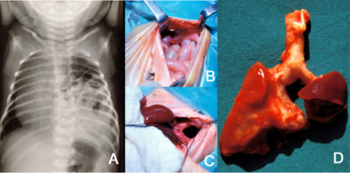
Tovar,JA,Universidad Autonoma de Madrid, Department of Pediatric Surgery, Hospital Universitario La Paz (Openi/National Library of Medicine) Source:https://openi.nlm.nih.gov/detailedresult?img=PMC3261088_1750-1172-7-1-1&query=WAGR%20Syndrome&it=xg&req=4&npos=52 Description:F1: A: Plain X-ray of the thorax of a newborn with CDH. There are bowel loops into the left hemi-thorax, the mediastinum is displaced to the contralateral side and the space occupied by the lung is reduced. B and C: At laparotomy, a left, posterolateral diaphragmatic hernia was discovered. In B, small bowel loops can be seen entering the thorax through the orifice. In C, this is seen after reducing the contents of the hernia. D: The patient died of severe persistent pulmonary hypertension days later. At autopsy, extreme left lung hypoplasia and less severe right lung hypoplasia were discovered.
File history
Click on a date/time to view the file as it appeared at that time.
| Date/Time | Thumbnail | Dimensions | User | Comment | |
|---|---|---|---|---|---|
| current | 15:11, 4 November 2021 | | 512 × 253 (329 KB) | Ozzie10aaaa (talk | contribs) | Tovar,JA,Universidad Autonoma de Madrid, Department of Pediatric Surgery, Hospital Universitario La Paz (Openi/National Library of Medicine) Source:https://openi.nlm.nih.gov/detailedresult?img=PMC3261088_1750-1172-7-1-1&query=WAGR%20Syndrome&it=xg&req=4&npos=52 Description:F1: A: Plain X-ray of the thorax of a newborn with CDH. There are bowel loops into the left hemi-thorax, the mediastinum is displaced to the contralateral side and the space occupied by the lung is reduced. B and C: At lap... |
You cannot overwrite this file.
File usage
There are no pages that use this file.