File:PMC3284108 IJMR-134-982-g001.png
PMC3284108_IJMR-134-982-g001.png (512 × 139 pixels, file size: 69 KB, MIME type: image/png)
License
Attribution-NonCommercial-ShareAlike 3.0 Unported (CC BY-NC-SA 3.0)
Summary
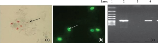
Author:Kulkarni S, Patsute S, Chandane M, Risbud A,National AIDS Research Institute , Naidu Municipal Corporation Hospital(Openi/National library of Medicine)Source:https://openi.nlm.nih.gov/detailedresult?img=PMC3284108_IJMR-134-982-g001&query=Microsporidiosis&it=xg&req=4&npos=4 Description:F1: (a) Stool smear stained with modified trichome stain showing microsporidial spores (arrow); (b) Stool smear stained by Uvitex 2B & examined with UV light. spores of microsporidia show typical elongated shape (arrow); (c) Agaraose gel electrophoresis of PCR product agarose gel electrophoresis of polymerase chain reaction (PCR product. Lane1: Marker 100 bp, Lane 2: positive sample, Lane 3: negative control and Lane 4: positive control).
File history
Click on a date/time to view the file as it appeared at that time.
| Date/Time | Thumbnail | Dimensions | User | Comment | |
|---|---|---|---|---|---|
| current | 23:44, 23 December 2021 | 512 × 139 (69 KB) | Ozzie10aaaa (talk | contribs) | Author:Kulkarni S, Patsute S, Chandane M, Risbud A,National AIDS Research Institute , Naidu Municipal Corporation Hospital(Openi/National library of Medicine)Source:https://openi.nlm.nih.gov/detailedresult?img=PMC3284108_IJMR-134-982-g001&query=Microsporidiosis&it=xg&req=4&npos=4 Description:F1: (a) Stool smear stained with modified trichome stain showing microsporidial spores (arrow); (b) Stool smear stained by Uvitex 2B & examined with UV light. spores of microsporidia show typical elongate... |
You cannot overwrite this file.
File usage
There are no pages that use this file.