File:PMC3309207 arm-35-436-g003.png
PMC3309207_arm-35-436-g003.png (471 × 196 pixels, file size: 178 KB, MIME type: image/png)
License
Attribution-NonCommercial 3.0 Unported (CC BY-NC 3.0)
Summary
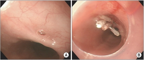
Author:Jung JH, Kim JS, Kim YK,Department of Physical Medicine and Rehabilitation, Kwandong University College of Medicine(Openi/National Library of Medicine) Source:https://openi.nlm.nih.gov/detailedresult?img=PMC3309207_arm-35-436-g003&query=Tracheoesophageal%20fistula&it=xg&req=4&npos=5 Description:F3: Esophagoduodenoscopic images: About the 3 mm-sized tracheoesophageal fistula was seen. And also the bubble from the trachea was observed. The fistula was located in the esophagus about 33 cm from the upper incisor (A). Post endoscopic clipping. Endoscopic clipping was done by using 4 clips (B).
File history
Click on a date/time to view the file as it appeared at that time.
| Date/Time | Thumbnail | Dimensions | User | Comment | |
|---|---|---|---|---|---|
| current | 23:03, 7 January 2022 | | 471 × 196 (178 KB) | Ozzie10aaaa (talk | contribs) | Author:Jung JH, Kim JS, Kim YK,Department of Physical Medicine and Rehabilitation, Kwandong University College of Medicine(Openi/National Library of Medicine) Source:https://openi.nlm.nih.gov/detailedresult?img=PMC3309207_arm-35-436-g003&query=Tracheoesophageal%20fistula&it=xg&req=4&npos=5 Description:F3: Esophagoduodenoscopic images: About the 3 mm-sized tracheoesophageal fistula was seen. And also the bubble from the trachea was observed. The fistula was located in the esophagus about 33 cm... |
You cannot overwrite this file.
File usage
There are no pages that use this file.