File:PMC3366251 RRP2012-258524.017.png
Jump to navigation
Jump to search
Size of this preview: 246 × 600 pixels. Other resolutions: 98 × 240 pixels | 420 × 1,024 pixels.
Original file (420 × 1,024 pixels, file size: 390 KB, MIME type: image/png)
Summary
| Description |
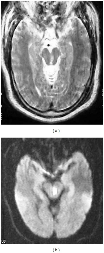
English: fig17: Claude Syndrome. Severely motion degraded axial T2-weighted (a) and DWI (b) images of the midbrain demonstrate a subacute infarct within the left midbrain tegmentum, with extension from the Sylvian aqueduct to the red nucleus. This patient presented with downward abduction of the left eye and right leg incoordination with poor gait. The visual disturbance is related to the involvement of the oculomotor nerve fascicles. Injury to the dentato-rubro fibers and/or red nucleus resulted in ataxia. |
| Date | |
| Source | https://openi.nlm.nih.gov/detailedresult?img=PMC3366251_RRP2012-258524.017&query=claude%27s%20syndrome&it=xg&req=4&npos=2 |
| Author | Ruchalski K, Hathout GM |
Licensing
English: This file is licensed CC BY-NC 4.0
This file was uploaded with UploadWizard.
File history
Click on a date/time to view the file as it appeared at that time.
| Date/Time | Thumbnail | Dimensions | User | Comment | |
|---|---|---|---|---|---|
| current | 19:39, 13 September 2022 | 420 × 1,024 (390 KB) | Ozzie10aaaa (talk | contribs) | Uploaded a work by Ruchalski K, Hathout GM from https://openi.nlm.nih.gov/detailedresult?img=PMC3366251_RRP2012-258524.017&query=claude%27s%20syndrome&it=xg&req=4&npos=2 with UploadWizard |
You cannot overwrite this file.
File usage
There are no pages that use this file.