File:PMC3400396 CRIM2012-121743.003.png

Original file (512 × 857 pixels, file size: 864 KB, MIME type: image/png)
License
Attribution 3.0 Unported (CC BY 3.0)
Summary
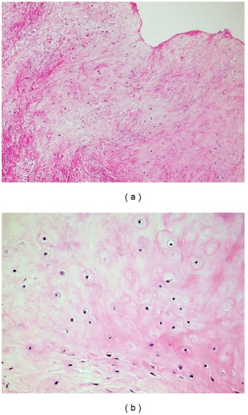
Author:Watanabe F, Saiki T, Ochochi Y, Department of Otolaryngology, Head and Neck Surgery, Steel Memorial Hirohata Hospital(Openi/National Library of Medicine) Source:https://openi.nlm.nih.gov/detailedresult?img=PMC3400396_CRIM2012-121743.003&query=Extraskeletal%20chondroma&it=xg&req=4&npos=17 Description:fig3: Histopathologically, (a) the tumor consists of hyaline cartilage and collagenous fibrous tissue (H&E stain, ×40). (b) The hyaline cartilaginous tissue contains homogeneous chondrocytes with round nuclei and chondrocytic lacunae (H&E stain, ×200).
File history
Click on a date/time to view the file as it appeared at that time.
| Date/Time | Thumbnail | Dimensions | User | Comment | |
|---|---|---|---|---|---|
| current | 21:59, 23 February 2022 |  | 512 × 857 (864 KB) | Ozzie10aaaa (talk | contribs) | Author:Watanabe F, Saiki T, Ochochi Y, Department of Otolaryngology, Head and Neck Surgery, Steel Memorial Hirohata Hospital(Openi/National Library of Medicine) Source:https://openi.nlm.nih.gov/detailedresult?img=PMC3400396_CRIM2012-121743.003&query=Extraskeletal%20chondroma&it=xg&req=4&npos=17 Description:fig3: Histopathologically, (a) the tumor consists of hyaline cartilage and collagenous fibrous tissue (H&E stain, ×40). (b) The hyaline cartilaginous tissue contains homogeneous chondrocyte... |
You cannot overwrite this file.
File usage
There are no pages that use this file.