File:PMC3414695 268 2012 1651 Fig2 HTML.png
Jump to navigation
Jump to search

Size of this preview: 359 × 600 pixels. Other resolutions: 144 × 240 pixels | 512 × 855 pixels.
Original file (512 × 855 pixels, file size: 632 KB, MIME type: image/png)
Summary
| Description |
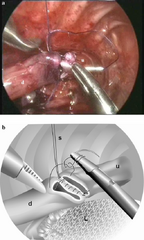
English: Fig2: Start of a running suture along the anterior side of the esophagus (this can be the same suture used for the posterior wall after it has been tied to the stay suture). Upper esophagus (u), distal esophagus (d), and lung (L), as well as the stay suture (S), are marked. b Drawing of the picture in a demonstrating the start of the anterior running suture |
| Date | |
| Source | https://openi.nlm.nih.gov/detailedresult?img=PMC3414695_268_2012_1651_Fig2_HTML&query=Esophageal%20atresia&it=xg&req=4&npos=9 |
| Author | van der Zee DC, Tytgat SH, Zwaveling S, van Herwaarden MY, Vieira-Travassos D |
Licensing
English: This file is licensed CC BY-NC 4.0
This file was uploaded with UploadWizard.
File history
Click on a date/time to view the file as it appeared at that time.
| Date/Time | Thumbnail | Dimensions | User | Comment | |
|---|---|---|---|---|---|
| current | 20:38, 2 January 2023 | | 512 × 855 (632 KB) | Ozzie10aaaa (talk | contribs) | Uploaded a work by van der Zee DC, Tytgat SH, Zwaveling S, van Herwaarden MY, Vieira-Travassos D from https://openi.nlm.nih.gov/detailedresult?img=PMC3414695_268_2012_1651_Fig2_HTML&query=Esophageal%20atresia&it=xg&req=4&npos=9 with UploadWizard |
You cannot overwrite this file.
File usage
There are no pages that use this file.