File:PMC3415789 rbhh-33-389-g01.png
Jump to navigation
Jump to search
No higher resolution available.
PMC3415789_rbhh-33-389-g01.png (512 × 165 pixels, file size: 167 KB, MIME type: image/png)
Summary
| Description |
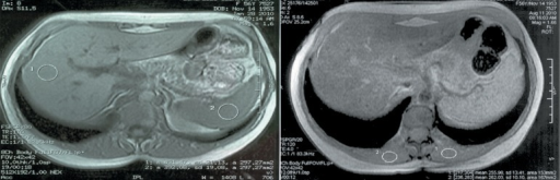
English: F1: Comparative study of upper abdomen MRI shows liver parenchyma with an increased signal in the left image due to increases in iron. On the right, after six months of treatment with deferasirox, a reduction in iron was observed by the MRI signal attenuation |
| Date | |
| Source | https://openi.nlm.nih.gov/detailedresult?img=PMC3415789_rbhh-33-389-g01&query=&req=4 |
| Author | Roberti Mdo R, Borges Filho HM, Gonçalves CH, Lima FL |
Licensing
English: This file is licensed CC BY-NC 3.0
This file was uploaded with UploadWizard.
File history
Click on a date/time to view the file as it appeared at that time.
| Date/Time | Thumbnail | Dimensions | User | Comment | |
|---|---|---|---|---|---|
| current | 21:55, 24 June 2022 | 512 × 165 (167 KB) | Ozzie10aaaa (talk | contribs) | Uploaded a work by Roberti Mdo R, Borges Filho HM, Gonçalves CH, Lima FL from https://openi.nlm.nih.gov/detailedresult?img=PMC3415789_rbhh-33-389-g01&query=&req=4 with UploadWizard |
You cannot overwrite this file.
File usage
There are no pages that use this file.