File:PMC3438271 kcj-42-575-g002.png
Jump to navigation
Jump to search
No higher resolution available.
PMC3438271_kcj-42-575-g002.png (512 × 379 pixels, file size: 192 KB, MIME type: image/png)
Summary
| Description |
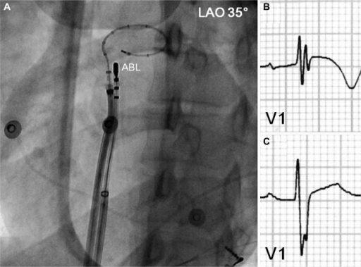
English: F2: Intracardiac mapping for monomorphic PVCs of RVOT origin. A: a deflectable ablation catheter and a 20-pole variable circular mapping catheter were placed at RVOT using long guiding sheathes. The circular mapping catheter with 25 mm diameter was positioned at 1 cm below pulmonary valve. The tip of ablation catheter was positioned at anterolateral wall of RVOT 2 cm below pulmonary valve, where the earliest ventricular activation signal was recorded during activation mapping for spontaneous PVCs. B: ECG before radiofrequency catheter ablation showed fragmented QRS complex with T wave inversion in V1. C: however, the fragmentation of QRS complex and T wave inversion disappeared after successful ablation. PVC: premature ventricular complex, RVOT: right ventricular outflow tract, ABL: ablation catheter, LAO: left anterior oblique view, ECG: eletrocardiogram. |
| Date | |
| Source | https://openi.nlm.nih.gov/detailedresult?img=PMC3438271_kcj-42-575-g002&query=Catheter%20ablation&it=xg&req=4&npos=2 |
| Author | Cho YR, Park JS |
Licensing
English: This file is licensed CC BY-NC 3.0
This file was uploaded with UploadWizard.
File history
Click on a date/time to view the file as it appeared at that time.
| Date/Time | Thumbnail | Dimensions | User | Comment | |
|---|---|---|---|---|---|
| current | 21:42, 12 October 2022 | | 512 × 379 (192 KB) | Ozzie10aaaa (talk | contribs) | Uploaded a work by Cho YR, Park JS from https://openi.nlm.nih.gov/detailedresult?img=PMC3438271_kcj-42-575-g002&query=Catheter%20ablation&it=xg&req=4&npos=2 with UploadWizard |
You cannot overwrite this file.
File usage
There are no pages that use this file.