File:PMC3471639 12-0621-F1.png
Jump to navigation
Jump to search
Size of this preview: 281 × 599 pixels. Other resolutions: 112 × 240 pixels | 480 × 1,024 pixels.
Original file (480 × 1,024 pixels, file size: 357 KB, MIME type: image/png)
Summary
| Description |
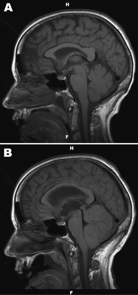
English: F1: A) Noncontrast, sagittal T1-weighted magnetic resonance image of the brain of a 67-year-old woman with suspected Powassan virus encephalitis, obtained 4 days after admission. Image is notable for nonspecific signal changes within the thalami, midbrain, cerebellar vermis, and both cerebellar hemispheres. B) Noncontrast, sagittal T1-weighted magnetic resonance image of the brain obtained 8 days after patient’s admission. Changes include marked interval progression of signal abnormality involving the cerebellum, thalamus, and midbrain. Mass effect within the posterior fossa and crowding of structures at the foramen magnum are also evident. Marked dilatation of the lateral and third ventricles with acute hydrocephalus is apparent. |
| Date | |
| Source | https://openi.nlm.nih.gov/detailedresult?img=PMC3471639_12-0621-F1&query=Powassan%20encephalitis&it=xg&req=4&npos=4 |
| Author | Birge J, Sonnesyn S |
Licensing
{{subst:Custom license marker added by UW}} https://creativecommons.org/publicdomain/zero/1.0/ CC0 1.0 Universal (CC0 1.0) Public Domain Dedication
This file was uploaded with UploadWizard.
File history
Click on a date/time to view the file as it appeared at that time.
| Date/Time | Thumbnail | Dimensions | User | Comment | |
|---|---|---|---|---|---|
| current | 21:30, 30 October 2022 | | 480 × 1,024 (357 KB) | Ozzie10aaaa (talk | contribs) | Uploaded a work by Birge J, Sonnesyn S from https://openi.nlm.nih.gov/detailedresult?img=PMC3471639_12-0621-F1&query=Powassan%20encephalitis&it=xg&req=4&npos=4 with UploadWizard |
You cannot overwrite this file.
File usage
There are no pages that use this file.