File:PMC3492682 jkms-27-1428-g003.png
Jump to navigation
Jump to search
No higher resolution available.
PMC3492682_jkms-27-1428-g003.png (512 × 168 pixels, file size: 204 KB, MIME type: image/png)
Summary
| Description |
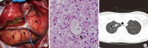
English: F3: Surgical and pathologic findings (August 2011). (A) An operative photograph shows a hemorrhagic cyst located in the middle frontal gyrus just anterior to the precentral gyrus (arrows). (B) Hematoxylin and eosin staining reveals an egg of Paragonimus (arrow) along with granulomatous inflammation (× 200). Note the thick asymmetric shell with a flattened side (arrow). (C) A chest CT scan (September 2011) reveals conglomerated, thin-walled cystic lesions with a nodule at the apex of the right upper lobe (arrow). |
| Date | |
| Source | https://openi.nlm.nih.gov/detailedresult?img=PMC3492682_jkms-27-1428-g003&query=&req=4 |
| Author | Koh EJ, Kim SK, Wang KC, Chai JY, Chong S, Park SH, Cheon JE, Phi JHf |
Licensing
English: This file is licensed CC BY-NC 3.0
This file was uploaded with UploadWizard.
File history
Click on a date/time to view the file as it appeared at that time.
| Date/Time | Thumbnail | Dimensions | User | Comment | |
|---|---|---|---|---|---|
| current | 22:46, 31 January 2023 | 512 × 168 (204 KB) | Ozzie10aaaa (talk | contribs) | Uploaded a work by Koh EJ, Kim SK, Wang KC, Chai JY, Chong S, Park SH, Cheon JE, Phi JHf from https://openi.nlm.nih.gov/detailedresult?img=PMC3492682_jkms-27-1428-g003&query=&req=4 with UploadWizard |
You cannot overwrite this file.
File usage
There are no pages that use this file.