File:PMC3492686 jkms-27-1444-g002.png
PMC3492686_jkms-27-1444-g002.png (512 × 254 pixels, file size: 296 KB, MIME type: image/png)
License
Attribution-NonCommercial 3.0 Unported (CC BY-NC 3.0)
Summary
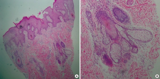
Author:Oh GN, Kim JY, Choi JE, Ahn HH, Kye YC, Seo SH ,Department of Dermatology, Korea University College of Medicine (Openi/National Library of Medicine)Source:https://openi.nlm.nih.gov/detailedresult?img=PMC3492686_jkms-27-1444-g002&query=Phakomatosis%20pigmentokeratotica&it=xg&req=4&npos=3 Description:F2: Histopathology of the skin lesion. (A) A skin biopsy from the right side of the cheek showed hyperkeratosis, moderate papillomatosis and acanthosis. There was a substantial increase in the number of mature sebaceous glands, hair follicles and eccrine glands in the epidermis (H&E stain, × 40). (B) At high power magnification, immature and inceased sebaceous glands were observed (H&E stain, × 100).
File history
Click on a date/time to view the file as it appeared at that time.
| Date/Time | Thumbnail | Dimensions | User | Comment | |
|---|---|---|---|---|---|
| current | 17:54, 11 April 2022 | | 512 × 254 (296 KB) | Ozzie10aaaa (talk | contribs) | Author:Oh GN, Kim JY, Choi JE, Ahn HH, Kye YC, Seo SH ,Department of Dermatology, Korea University College of Medicine (Openi/National Library of Medicine)Source:https://openi.nlm.nih.gov/detailedresult?img=PMC3492686_jkms-27-1444-g002&query=Phakomatosis%20pigmentokeratotica&it=xg&req=4&npos=3 Description:F2: Histopathology of the skin lesion. (A) A skin biopsy from the right side of the cheek showed hyperkeratosis, moderate papillomatosis and acanthosis. There was a substantial increase in t... |
You cannot overwrite this file.
File usage
There are no pages that use this file.