File:PMC3546075 pone.0053653.jpg
Jump to navigation
Jump to search
Size of this preview: 489 × 600 pixels. Other resolutions: 196 × 240 pixels | 512 × 628 pixels.
Original file (512 × 628 pixels, file size: 63 KB, MIME type: image/jpeg)
Summary
| Description |
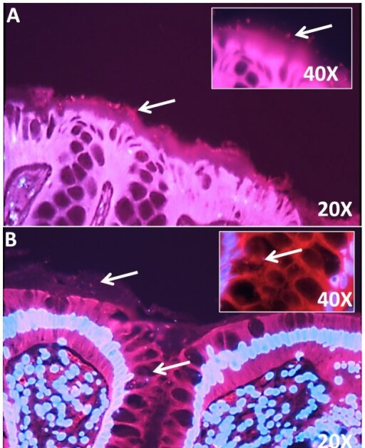
English: pone-0053653-g002: Representative fluorescence in situ hybridization targeting Fusobacterium sp. in colorectal mucosal biopsy sections using bacterial 16S rRNA probes.Fig. 2A–B are composite images of Cy3 and DAPI views of sections hybridized with a Fusobacterium-specific probe. Fusobacterium species is localized within the mucus layer of colorectal sections (A) 20X and 40X. Fusobacterium species is localized within the crypts of colorectal section (B) 20X and 40X. Fig. 2C (20X and 40X) is a positive control and shows sections stained with general bacteria probe (Eub 388). General bacteria, including most Eubacteria species, are localized to the mucus layer above the epithelium. White arrows point to bacteria either in mucus layer above the colonic epithelium or within the crypt. |
| Date | |
| Source | https://openi.nlm.nih.gov/detailedresult?img=PMC3546075_pone.0053653.g002&query=Fusobacterium&it=xg&req=4&npos=1 |
| Author | McCoy AN, Araújo-Pérez F, Azcárate-Peril A, Yeh JJ, Sandler RS, Keku TO |
Licensing
English: This file is licensed CC BY-NC 4.0
This file was uploaded with UploadWizard.
File history
Click on a date/time to view the file as it appeared at that time.
| Date/Time | Thumbnail | Dimensions | User | Comment | |
|---|---|---|---|---|---|
| current | 18:30, 16 June 2023 | | 512 × 628 (63 KB) | Ozzie10aaaa (talk | contribs) | Uploaded a work by McCoy AN, Araújo-Pérez F, Azcárate-Peril A, Yeh JJ, Sandler RS, Keku TO from https://openi.nlm.nih.gov/detailedresult?img=PMC3546075_pone.0053653.g002&query=Fusobacterium&it=xg&req=4&npos=1 with UploadWizard |
You cannot overwrite this file.
File usage
There are no pages that use this file.