File:PMC3573807 cro-0006-0055-g03.png
PMC3573807_cro-0006-0055-g03.png (512 × 380 pixels, file size: 429 KB, MIME type: image/png)
License
Attribution-NonCommercial-NoDerivs 3.0 Unported (CC BY-NC-ND 3.0)
- &
Summary
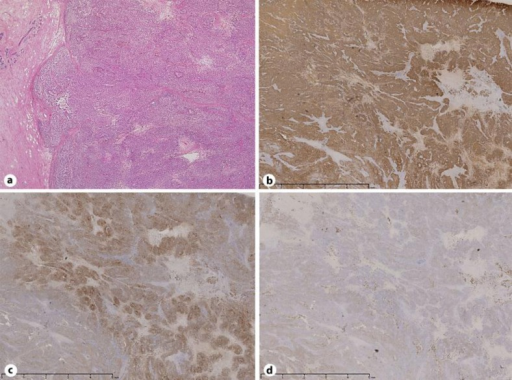
Author:Fukada I, Nishimura S, Tanabe M, Morizono H, Makita M, Gomi N, Horii R, Akiyama F, Iwase T,Department of Breast Medical Oncology, the Cancer Institute Hospital of the Japanese Foundation for Cancer Research, and the Cancer Institute of the Japanese Foundation for Cancer Research(Openi/National Library of Medicine) Source:https://openi.nlm.nih.gov/detailedresult?img=PMC3573807_cro-0006-0055-g03&query=&req=4 Description:F3: Histopathological findings. A well-defined solid mass comprising nests of small, spindle-shaped, melanin-containing cells was observed (a). The tumor cells were immunohistochemically positive for HMB45 (b), S-100 (c), and Melan-A (d).
File history
Click on a date/time to view the file as it appeared at that time.
| Date/Time | Thumbnail | Dimensions | User | Comment | |
|---|---|---|---|---|---|
| current | 22:45, 16 March 2022 | | 512 × 380 (429 KB) | Ozzie10aaaa (talk | contribs) | Author:Fukada I, Nishimura S, Tanabe M, Morizono H, Makita M, Gomi N, Horii R, Akiyama F, Iwase T,Department of Breast Medical Oncology, the Cancer Institute Hospital of the Japanese Foundation for Cancer Research, and the Cancer Institute of the Japanese Foundation for Cancer Research(Openi/National Library of Medicine) Source:https://openi.nlm.nih.gov/detailedresult?img=PMC3573807_cro-0006-0055-g03&query=&req=4 Description:F3: Histopathological findings. A well-defined solid mass comprising... |
You cannot overwrite this file.
File usage
There are no pages that use this file.