File:PMC3619661 jcnsd-4-2012-073f1 (1).png
Jump to navigation
Jump to search
No higher resolution available.
PMC3619661_jcnsd-4-2012-073f1_(1).png (512 × 254 pixels, file size: 92 KB, MIME type: image/png)
Summary
| Description |
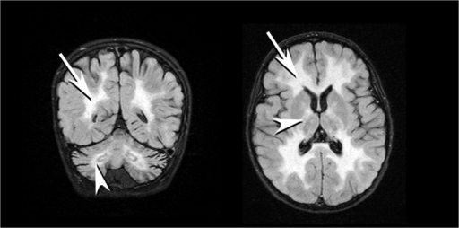
English: f1-jcnsd-4-2012-073: Sample MR images from our study cohort showing advanced leukodystrophy.Notes: Left: case #1, coronal FLAIR images obtained at the level of posterior fossa demonstrating confluent areas of white matter degeneration in the periventicular region (long arrow) as well as the cerebellum (arroe head). Right: case #2, axial FLAIR images obtained at the level of the basal ganglia showing confluent areas of signal abnormality in the sub-cortical regions (long arrow) as well as the internal capsule (arrow head). |
| Date | |
| Source | https://openi.nlm.nih.gov/detailedresult?img=PMC3619661_jcnsd-4-2012-073f1&query=Leukodystrophy&it=xg&req=4&npos=7 |
| Author | Assadi M, Wang DJ, Anderson K, Carran M, Bilaniuk L, Leone P |
Licensing
English: This file is licensed CC BY-NC 3.0
This file was uploaded with UploadWizard.
File history
Click on a date/time to view the file as it appeared at that time.
| Date/Time | Thumbnail | Dimensions | User | Comment | |
|---|---|---|---|---|---|
| current | 21:07, 14 October 2023 | | 512 × 254 (92 KB) | Ozzie10aaaa (talk | contribs) | Uploaded a work by Assadi M, Wang DJ, Anderson K, Carran M, Bilaniuk L, Leone P from https://openi.nlm.nih.gov/detailedresult?img=PMC3619661_jcnsd-4-2012-073f1&query=Leukodystrophy&it=xg&req=4&npos=7 with UploadWizard |
You cannot overwrite this file.
File usage
There are no pages that use this file.
.png&oldid=1254091)