File:PMC3654851 mv-v19-935-f4 (1).png
PMC3654851_mv-v19-935-f4_(1).png (512 × 282 pixels, file size: 243 KB, MIME type: image/png)
License
Attribution 3.0 Unported (CC BY 3.0)
Summary
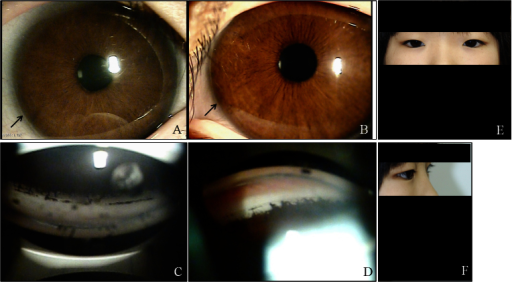
Author:Kim GN, Ki CS, Seo SW, Yoo JM, Han YS, Chung IY, Park JM, Kim SJ ,Department of Ophthalmology, Gyeongsang National University, College of Medicine (Openi/National Library of Medicine) Source:https://openi.nlm.nih.gov/detailedresult?img=PMC3654851_mv-v19-935-f4&query=Axenfeld%E2%80%93Rieger%20syndrome&it=xg&req=4&npos=20 Description:f4: Ocular characteristics and systemic anomalies of patient II:2. Slit lamp photographs showed iris hypoplasia and posterior embryotoxon (black arrows) in both eyes, but Haab’s striae were observed only in the right eye (A, B). Gonioscopy showed open angles in both eyes with anterior insertion of the iris into the trabecular meshwork, prominent iris processes, and broad-based synechiae at places in both eyes (C, D). Physical examination revealed hypertelorism, telecanthus, a flat face, and a flat broad nasal bridge (E, F).
File history
Click on a date/time to view the file as it appeared at that time.
| Date/Time | Thumbnail | Dimensions | User | Comment | |
|---|---|---|---|---|---|
| current | 17:55, 24 September 2021 | | 512 × 282 (243 KB) | Ozzie10aaaa (talk | contribs) | Author:Kim GN, Ki CS, Seo SW, Yoo JM, Han YS, Chung IY, Park JM, Kim SJ ,Department of Ophthalmology, Gyeongsang National University, College of Medicine (Openi/National Library of Medicine) Source:https://openi.nlm.nih.gov/detailedresult?img=PMC3654851_mv-v19-935-f4&query=Axenfeld%E2%80%93Rieger%20syndrome&it=xg&req=4&npos=20 Description:f4: Ocular characteristics and systemic anomalies of patient II:2. Slit lamp photographs showed iris hypoplasia and posterior embryotoxon (black arrows) in... |
You cannot overwrite this file.
File usage
There are no pages that use this file.
.png&oldid=1236498)