File:PMC3656102 ppat.1003374.png
Jump to navigation
Jump to search
No higher resolution available.
PMC3656102_ppat.1003374.png (160 × 216 pixels, file size: 82 KB, MIME type: image/png)
Summary
| Description |
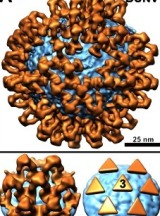
English: ppat-1003374-g006: Structural diversity of bunyavirus surface glycoprotein architectures.(A–C) Structures of Gn–Gc glycoprotein spikes (orange) are shown mapped onto the membrane surface (cyan) of: (A) an orthobunyavirus (Bunyamwera virus; BUNV), (B) a hantavirus (Tula virus; TULV; EMD-170411), and (C) a phlebovirus (Rift Valley fever virus; RVFV; EMD-15507). At the bottom of each panel, we show a close-up view of a glycoprotein spike cluster (left) and a schematic representation of the spike arrangement (right). Symmetries of individual spikes are annotated. |
| Date | |
| Source | https://openi.nlm.nih.gov/detailedresult?img=PMC3656102_ppat.1003374.g006&query=Bunyamwera%20orthobunyavirus&it=xg&req=4&npos=10 |
| Author | Bowden TA, Bitto D, McLees A, Yeromonahos C, Elliott RM, Huiskonen JT |
Licensing
English: This file is licensed CC BY-NC 4.0
This file was uploaded with UploadWizard.
File history
Click on a date/time to view the file as it appeared at that time.
| Date/Time | Thumbnail | Dimensions | User | Comment | |
|---|---|---|---|---|---|
| current | 02:03, 28 March 2023 | | 160 × 216 (82 KB) | Ozzie10aaaa (talk | contribs) | Cropped 8 % horizontally, 13 % vertically using CropTool with precise mode. |
You cannot overwrite this file.
File usage
There are no pages that use this file.