File:PMC3665880 aps-40-277-g002.png
PMC3665880_aps-40-277-g002.png (512 × 338 pixels, file size: 392 KB, MIME type: image/png)
License
Attribution-NonCommercial 3.0 Unported (CC BY-NC 3.0)
Summary
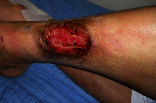
Author:Seok HH, Noh Y, Jeong EC, Park JU, Hong YH,Department of Plastic and Reconstructive Surgery, Seoul National University College of Medicine, Seoul, Korea (Openi/National Library of Medicine) Source:https://openi.nlm.nih.gov/detailedresult?img=PMC3665880_aps-40-277-g002&query=Sneddon%27s%20syndrome&it=xg&req=4&npos=2 Description:F2: The ulcers with livedo reticularis. This photograph shows the wound on the right pretibial area. After the second debridement, this ulcer grossly showed relatively healthy granulation tissue without any sign of infection.
File history
Click on a date/time to view the file as it appeared at that time.
| Date/Time | Thumbnail | Dimensions | User | Comment | |
|---|---|---|---|---|---|
| current | 20:01, 4 August 2021 | | 512 × 338 (392 KB) | Ozzie10aaaa (talk | contribs) | Author:Seok HH, Noh Y, Jeong EC, Park JU, Hong YH,Department of Plastic and Reconstructive Surgery, Seoul National University College of Medicine, Seoul, Korea (Openi/National Library of Medicine) Source:https://openi.nlm.nih.gov/detailedresult?img=PMC3665880_aps-40-277-g002&query=Sneddon%27s%20syndrome&it=xg&req=4&npos=2 Description:F2: The ulcers with livedo reticularis. This photograph shows the wound on the right pretibial area. After the second debridement, this ulcer grossly showed rela... |
You cannot overwrite this file.
File usage
There are no pages that use this file.