File:PMC3713254 234 2013 1180 Fig3 HTML.png
Jump to navigation
Jump to search

Size of this preview: 395 × 599 pixels. Other resolutions: 158 × 240 pixels | 512 × 777 pixels.
Original file (512 × 777 pixels, file size: 620 KB, MIME type: image/png)
Summary
| Description |
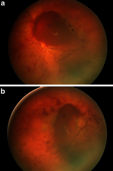
English: Fig3: RetCam pictures of patient 6. a Right fundus. There are white-centered superficial intraretinal hemorrhages (squares) and deep intraretinal hemorrhages (arrowheads). There is a large preretinal hemorrhage obscuring part of the optic disk (circle). b Left fundus. There are some superficial intraretinal hemorrhages (squares) and less obvious deep intraretinal hemorrhages. There is a larger preretinal hemorrhage obscuring the optic disk (circle). The suggestion of some schitic retinal changes is felt to represent an artifact of the imaging system (arrows) |
| Date | |
| Source | https://openi.nlm.nih.gov/detailedresult?img=PMC3713254_234_2013_1180_Fig3_HTML&query=Retinal%20haemorrhage&it=xg&req=4&npos=3 |
| Author | Zuccoli G, Panigrahy A, Haldipur A, Willaman D, Squires J, Wolford J, Sylvester C, Mitchell E, Lope LA, Nischal KK, Berger RP |
Licensing
English: This file is licensed CC BY-NC 4.0
This file was uploaded with UploadWizard.
File history
Click on a date/time to view the file as it appeared at that time.
| Date/Time | Thumbnail | Dimensions | User | Comment | |
|---|---|---|---|---|---|
| current | 19:43, 14 July 2022 | | 512 × 777 (620 KB) | Ozzie10aaaa (talk | contribs) | Uploaded a work by Zuccoli G, Panigrahy A, Haldipur A, Willaman D, Squires J, Wolford J, Sylvester C, Mitchell E, Lope LA, Nischal KK, Berger RP from https://openi.nlm.nih.gov/detailedresult?img=PMC3713254_234_2013_1180_Fig3_HTML&query=Retinal%20haemorrhage&it=xg&req=4&npos=3 with UploadWizard |
You cannot overwrite this file.
File usage
There are no pages that use this file.