File:PMC3713982 12-1820-F.png
Jump to navigation
Jump to search
Size of this preview: 372 × 599 pixels. Other resolutions: 149 × 240 pixels | 512 × 824 pixels.
Original file (512 × 824 pixels, file size: 653 KB, MIME type: image/png)
Summary
| Description |
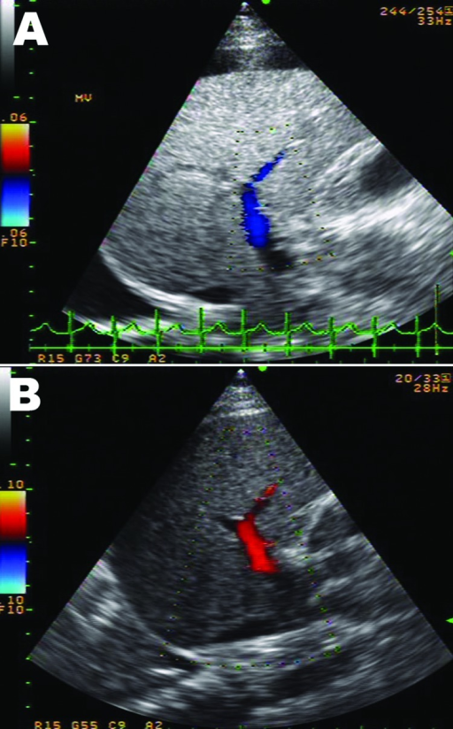
English: F1: Ultrasonograph with Doppler image of the liver of a 10-year-old boy with liver failure associated with dengue virus infection. A) Day 2 of hospitalization, showing reversed direction of blood flow in the right branch of the portal vein (hepatofugal flow). There was diffuse increased liver parenchymal echo, swelling of the gallbladder wall, and right pleural effusion. B) Day 5 of hospitalization, showing returning normal direction of portal venous flow (hepatopetal flow). Liver parenchymal echo changed to normal. Pleural fluid and swelling of the gallbladder wall also disappeared. |
| Date | |
| Source | https://openi.nlm.nih.gov/detailedresult?img=PMC3713982_12-1820-F&query=liver%20failure&it=xg&req=4&npos=6 |
| Author | Khongphatthanayothin A, Mahayosnond A, Poovorawan Y |
Licensing
{{subst:Custom license marker added by UW}} https://creativecommons.org/publicdomain/zero/1.0/ CC0 1.0 Universal (CC0 1.0) Public Domain Dedication
This file was uploaded with UploadWizard.
File history
Click on a date/time to view the file as it appeared at that time.
| Date/Time | Thumbnail | Dimensions | User | Comment | |
|---|---|---|---|---|---|
| current | 19:35, 31 July 2022 | | 512 × 824 (653 KB) | Ozzie10aaaa (talk | contribs) | Uploaded a work by Khongphatthanayothin A, Mahayosnond A, Poovorawan Y from https://openi.nlm.nih.gov/detailedresult?img=PMC3713982_12-1820-F&query=liver%20failure&it=xg&req=4&npos=6 with UploadWizard |
You cannot overwrite this file.
File usage
There are no pages that use this file.