File:PMC3777705 gr1.png
Jump to navigation
Jump to search
No higher resolution available.
PMC3777705_gr1.png (512 × 221 pixels, file size: 122 KB, MIME type: image/png)
Summary
| Description |
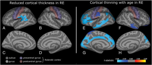
English: f0005: Cortical abnormalities in rolandic epilepsy.Inflated brain visualizations of the regions showing abnormal cortical morphology in RE. Subfigures A–D display reduced cortical thickness in RE (age and gender corrected), subfigures E–H depict regions showing cortical thinning for increasing age, an effect which was only found in the patients (gender corrected). Reduced cortical thickness was found predominantly in the supramarginal gyrus and partly covered the bank of the superior temporal sulcus, the superior temporal gyrus and the lower postcentral gyrus (A). Cortical thinning with age was predominantly found in the left hemisphere and involved the inferior frontal gyrus, the inferior postcentral and the supramarginal gyrus and the middle temporal gyri at the lateral side (E), and the cuneus, precuneus and cingulate cortex medially (G). |
| Date | |
| Source | https://openi.nlm.nih.gov/detailedresult?img=PMC3777705_gr1&query=Rolandic%20epilepsy&it=xg&req=4&npos=1 |
| Author | Overvliet GM, Besseling RM, Jansen JF, van der Kruijs SJ, Vles JS, Hofman PA, Ebus SC, de Louw A, Aldenkamp AP, Backes WH |
Licensing
English: This file is licensed CC BY-NC-SA 3.0
This file was uploaded with UploadWizard.
File history
Click on a date/time to view the file as it appeared at that time.
| Date/Time | Thumbnail | Dimensions | User | Comment | |
|---|---|---|---|---|---|
| current | 20:31, 19 June 2022 | | 512 × 221 (122 KB) | Ozzie10aaaa (talk | contribs) | Uploaded a work by Overvliet GM, Besseling RM, Jansen JF, van der Kruijs SJ, Vles JS, Hofman PA, Ebus SC, de Louw A, Aldenkamp AP, Backes WH from https://openi.nlm.nih.gov/detailedresult?img=PMC3777705_gr1&query=Rolandic%20epilepsy&it=xg&req=4&npos=1 with UploadWizard |
You cannot overwrite this file.
File usage
There are no pages that use this file.