File:PMC3834874 1752-1947-7-255-1.png

Original file (512 × 625 pixels, file size: 209 KB, MIME type: image/png)
License
Attribution 2.0 Generic (CC BY 2.0)
Summary
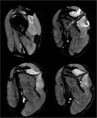
Author:Kumar I, Verma A, Srivastava A, Shukla RC,Department of Radiodiagnosis and Imaging, Institute of Medical Sciences, Banaras Hindu University(Openi/National Library of Medicine) Source:https://openi.nlm.nih.gov/detailedresult?img=PMC3834874_1752-1947-7-255-1&query=Parsonage%20Turner%20syndrome&it=xg&req=4&npos=3 Description:F1: Magnetic resonance imaging of patient 1 at the level of shoulder girdle including the periscapular muscles. Sagittal T2-weighted fat-suppressed contiguous section at the level of shoulder showing edema in suprasinatus (solid arrow) and infraspinatus (curved arrow) muscles, respectively above and below the spine of scapula (straight arrow). The brachial plexus in this patient, however did not reveal any abnormality.
File history
Click on a date/time to view the file as it appeared at that time.
| Date/Time | Thumbnail | Dimensions | User | Comment | |
|---|---|---|---|---|---|
| current | 22:51, 15 January 2022 | | 512 × 625 (209 KB) | Ozzie10aaaa (talk | contribs) | Author:Kumar I, Verma A, Srivastava A, Shukla RC,Department of Radiodiagnosis and Imaging, Institute of Medical Sciences, Banaras Hindu University(Openi/National Library of Medicine) Source:https://openi.nlm.nih.gov/detailedresult?img=PMC3834874_1752-1947-7-255-1&query=Parsonage%20Turner%20syndrome&it=xg&req=4&npos=3 Description:F1: Magnetic resonance imaging of patient 1 at the level of shoulder girdle including the periscapular muscles. Sagittal T2-weighted fat-suppressed contiguous section... |
You cannot overwrite this file.
File usage
There are no pages that use this file.