File:PMC3843328 IJRI-23-212-g005.png
PMC3843328_IJRI-23-212-g005.png (512 × 143 pixels, file size: 151 KB, MIME type: image/png)
License
Attribution-NonCommercial-ShareAlike 3.0 Unported (CC BY-NC-SA 3.0)
Summary
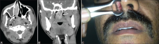
Author:Prabhu SM, Irodi A, Khiangte HL, Rupa V, Naina P, Department of Radiology, Christian Medical College, Vellore (Openi/National Library of Medicine) Source:https://openi.nlm.nih.gov/detailedresult?img=PMC3843328_IJRI-23-212-g005&query=Rhinosporidiosis&it=xg&req=4&npos=18 Description:F3: A 38-year-old male with epistaxis and nasal mass. Contrast-enhanced CT PNS (A) Axial image shows an enhancing soft tissue lesion (black arrow) in the right inferior nasal cavity extending through the choana into the nasopharynx. (B) Coronal section shows lobulated nasopharyngeal extension of the lesion (white arrow) with prominent leash of blood vessels. (C) Anterior rhinoscopy shows a red fleshy mass with whitish spots in the right nasal cavity. Diagnosis after surgery confirmed as rhinosporidiosis
File history
Click on a date/time to view the file as it appeared at that time.
| Date/Time | Thumbnail | Dimensions | User | Comment | |
|---|---|---|---|---|---|
| current | 19:12, 13 September 2021 | 512 × 143 (151 KB) | Ozzie10aaaa (talk | contribs) | Author:Prabhu SM, Irodi A, Khiangte HL, Rupa V, Naina P, Department of Radiology, Christian Medical College, Vellore (Openi/National Library of Medicine) Source:https://openi.nlm.nih.gov/detailedresult?img=PMC3843328_IJRI-23-212-g005&query=Rhinosporidiosis&it=xg&req=4&npos=18 Description:F3: A 38-year-old male with epistaxis and nasal mass. Contrast-enhanced CT PNS (A) Axial image shows an enhancing soft tissue lesion (black arrow) in the right inferior nasal cavity extending through the choa... |
You cannot overwrite this file.
File usage
There are no pages that use this file.