File:PMC3849310 kjo-27-454-g001.png
PMC3849310_kjo-27-454-g001.png (512 × 388 pixels, file size: 335 KB, MIME type: image/png)
License
Attribution-NonCommercial 3.0 Unported (CC BY-NC 3.0)
Summary
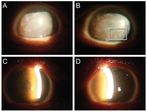
Author:Lee YK, Chang DJ, Chung SK.Department of Ophthalmology, The Catholic University of Korea College of Medicine (Openi/National Library of Medicine) Source:https://openi.nlm.nih.gov/detailedresult?img=PMC3849310_kjo-27-454-g001&query=Macular%20corneal%20dystrophy&it=xg&req=4&npos=7 Description: F1: Slit lamp photography of the patient (A,B). (A) Right eye and (B) left eye. Diffusely hazy corneas with bilateral opacities were observed. There were multiple irregular, grayish-white, dense, poorly delineated spots in the stroma. Slit lamp photography of the patient's son (C,D). (C) Right eye and (D) left eye. Haziness of the stroma with diffuse opacities was observed in both eyes. These findings were similar, but less severe in appearance compared to his mother's examination.
File history
Click on a date/time to view the file as it appeared at that time.
| Date/Time | Thumbnail | Dimensions | User | Comment | |
|---|---|---|---|---|---|
| current | 18:47, 12 August 2021 | | 512 × 388 (335 KB) | Ozzie10aaaa (talk | contribs) | Author:Lee YK, Chang DJ, Chung SK.Department of Ophthalmology, The Catholic University of Korea College of Medicine (Openi/National Library of Medicine) Source:https://openi.nlm.nih.gov/detailedresult?img=PMC3849310_kjo-27-454-g001&query=Macular%20corneal%20dystrophy&it=xg&req=4&npos=7 Description: F1: Slit lamp photography of the patient (A,B). (A) Right eye and (B) left eye. Diffusely hazy corneas with bilateral opacities were observed. There were multiple irregular, grayish-white, dense, p... |
You cannot overwrite this file.
File usage
There are no pages that use this file.